J Korean Soc Radiol.
2018 Sep;79(3):129-138. 10.3348/jksr.2018.79.3.129.
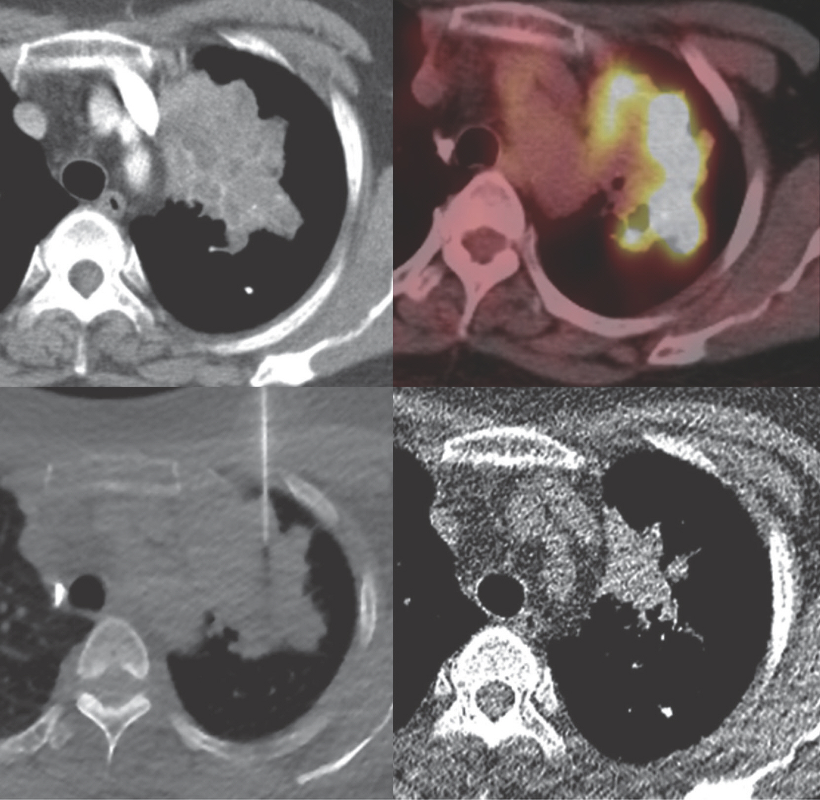
Study of the Efficacy of PET/CT in Lung Aspiration Biopsy and Factors Associated with False-Negative Results
- Affiliations
-
- 1Department of Radiology, Pusan National University Hospital, Busan, Korea. rabkingdom@naver.com
- 2Medical Research Institute, Pusan National University Hospital, Busan, Korea.
- 3Department of Pathology, Pusan National University Hospital, Busan, Korea.
- KMID: 2418654
- DOI: http://doi.org/10.3348/jksr.2018.79.3.129
Abstract
- PURPOSE
We compared the outcomes of percutaneous transthoracic needle aspiration biopsy (PCNA) of lung masses in cases with and without prior positron emission tomography/computed tomography (PET/CT) information, and investigated the factors associated with false-negative pathological results.
MATERIALS AND METHODS
From a total of 291 patients, 161 underwent PCNA without prior PET/CT imaging, while 130 underwent PET/CT before PCNA. Clinical characteristics, procedural variables, pathological results, and diagnostic success rates were compared between the 2 groups. Among patients with initial negative (non-specific benign) PCNA results, the radiological findings of these groups were compared to evaluate the predictors of false-negative lesions.
RESULTS
No significant difference was found in the clinical characteristics, procedural characteristics, and pathological results of the 2 groups, nor was the diagnostic rate significantly different between them (p = 0.818). Among patients with initial negative PCNA results, radiological characteristics were similar in both the groups. In multivariate analysis, the presence of necrosis (p = 0.005) and ground-glass opacity (GGO) (p = 0.011) were the significant characteristics that indicated an increased probability of initial false-negative results in PCNA.
CONCLUSION
Routine PET/CT did not have any additional benefit in patients undergoing PCNA of lung masses. The presence of necrosis or GGO could indicate an increased probability of false-negative pathological results.
MeSH Terms
Figure
Reference
-
1.National Lung Screening Trial Research Team. Church TR., Black WC., Aberle DR., Berg CD., Clingan KL, et al. Results of initial low-dose computed tomographic screening for lung cancer. N Engl J Med. 2013. 368:1980–1991.
Article2.Hoffman JM., Gambhir SS. Molecular imaging: the vision and opportunity for radiology in the future. Radiology. 2007. 244:39–47.
Article3.Bomanji JB., Costa DC., Ell PJ. Clinical role of positron emission tomography in oncology. Lancet Oncol. 2001. 2:157–164.
Article4.Cornelis F., Silk M., Schoder H., Takaki H., Durack JC., Erinjeri JP, et al. Performance of intra-procedural 18-fluorodeoxy-glucose PET/CT-guided biopsies for lesions suspected of malignancy but poorly visualized with other modalities. Eur J Nucl Med Mol Imaging. 2014. 41:2265–2272.
Article5.Klaeser B., Mueller MD., Schmid RA., Guevara C., Krause T., Wiskirchen J. PET-CT-guided interventions in the management of FDG-positive lesions in patients suffering from solid malignancies: initial experiences. Eur Radiol. 2009. 19:1780–1785.
Article6.Guralnik L., Rozenberg R., Frenkel A., Israel O., Keidar Z. Metabolic PET/CT-guided lung lesion biopsies: impact on diagnostic accuracy and rate of sampling error. J Nucl Med. 2015. 56:518–522.
Article7.Purandare NC., Kulkarni AV., Kulkarni SS., Roy D., Agrawal A., Shah S, et al. 18F-FDG PET/CT-directed biopsy: does it offer incremental benefit? Nucl Med Commun. 2013. 34:203–210.8.Stattaus J., Kuehl H., Ladd S., Schroeder T., Antoch G., Baba HA, et al. CT-guided biopsy of small liver lesions: visibility, artifacts, and corresponding diagnostic accuracy. Cardiovasc Intervent Radiol. 2007. 30:928–935.
Article9.Gelbman BD., Cham MD., Kim W., Libby DM., Smith JP., Port JL, et al. Radiographic and clinical characterization of false negative results from CT-guided needle biopsies of lung nodules. J Thorac Oncol. 2012. 7:815–820.
Article10.Hiraki T., Mimura H., Gobara H., Iguchi T., Fujiwara H., Sakurai J, et al. CT fluoroscopy-guided biopsy of 1,000 pulmonary lesions performed with 20-gauge coaxial cutting needles: diagnostic yield and risk factors for diagnostic failure. Chest. 2009. 136:1612–1617.11.Hansell DM., Bankier AA., MacMahon H., McLoud TC., Müller NL., Remy J. Fleischner Society: glossary of terms for thoracic imaging. Radiology. 2008. 246:697–722.
Article12.de Geus-Oei LF., van der Heijden HF., Visser EP., Hermsen R., van Hoorn BA., Timmer-Bonte JN, et al. Chemotherapy response evaluation with 18F-FDG PET in patients with nonsmall cell lung cancer. J Nucl Med. 2007. 48:1592–1598.
Article13.Hicks RJ., Kalff V., MacManus MP., Ware RE., Hogg A., McKenzie AF, et al. (18)F-FDG PET provides high-impact and powerful prognostic stratification in staging newly diagnosed nonsmall cell lung cancer. J Nucl Med. 2001. 42:1596–1604.14.Takeuchi S., Khiewvan B., Fox PS., Swisher SG., Rohren EM., Bassett RL Jr, et al. Impact of initial PET/CT staging in terms of clinical stage, management plan, and prognosis in 592 patients with nonsmall-cell lung cancer. Eur J Nucl Med Mol Imaging. 2014. 41:906–914.
Article15.Truong MT., Viswanathan C., Erasmus JJ. Positron emission tomography/computed tomography in lung cancer staging, prognosis, and assessment of therapeutic response. J Thorac Imaging. 2011. 26:132–146.
Article16.Cerci JJ., Pereira Neto CC., Krauzer C., Sakamoto DG., Vitola JV. The impact of coaxial core biopsy guided by FDG PET/CT in oncological patients. Eur J Nucl Med Mol Imaging. 2013. 40:98–103.
Article17.Klaeser B., Wiskirchen J., Wartenberg J., Weitzel T., Schmid RA., Mueller MD, et al. PET/CT-guided biopsies of metabolically active bone lesions: applications and clinical impact. Eur J Nucl Med Mol Imaging. 2010. 37:2027–2036.
Article18.Kim JI., Park CM., Kim H., Lee JH., Goo JM. Non-specific benign pathological results on transthoracic core-needle biopsy: how to differentiate false-negatives? Eur Radiol. 2017. 27:3888–3895.
Article19.Minot DM., Gilman EA., Aubry MC., Voss JS., Van Epps SG., Tuve DJ, et al. An investigation into false-negative transthoracic fine needle aspiration and core biopsy specimens. Diagn Cy-topathol. 2014. 42:1063–1068.
Article20.Tsukada H., Satou T., Iwashima A., Souma T. Diagnostic accuracy of CT-guided automated needle biopsy of lung nodules. AJR Am J Roentgenol. 2000. 175:239–243.
Article21.Yeow KM., Tsay PK., Cheung YC., Lui KW., Pan KT., Chou AS. Factors affecting diagnostic accuracy of CT-guided coaxial cutting needle lung biopsy: retrospective analysis of 631 procedures. J Vasc Interv Radiol. 2003. 14:581–588.
Article22.Heppner GH. Tumor heterogeneity. Cancer Res. 1984. 44:2259–2265.
Article23.Miles KA., Williams RE. Warburg revisited: imaging tumour blood flow and metabolism. Cancer Imaging. 2008. 8:81–86.
Article24.Swanton C. Intratumor heterogeneity: evolution through space and time. Cancer Res. 2012. 72:4875–4882.
Article25.Bar-Shalom R., Yefremov N., Guralnik L., Gaitini D., Frenkel A., Kuten A, et al. Clinical performance of PET/CT in evaluation of cancer: additional value for diagnostic imaging and patient management. J Nucl Med. 2003. 44:1200–1209.26.Kubota K. From tumor biology to clinical PET: a review of positron emission tomography (PET) in oncology. Ann Nucl Med. 2001. 15:471–486.
Article27.Hua Q., Zhu X., Zhang L., Zhao Y., Tang P., Ni J. Initial experience with real-time hybrid single-photon emission computed tomography/computed tomography-guided percutaneous transthoracic needle biopsy. Nucl Med Commun. 2017. 38:556–560.
Article28.Aoki T., Tomoda Y., Watanabe H., Nakata H., Kasai T., Hashimoto H, et al. Peripheral lung adenocarcinoma: correlation of thin-section CT findings with histologic prognostic factors and survival. Radiology. 2001. 220:803–809.
Article29.Lee HY., Lee KS. Ground-glass opacity nodules: histopathology, imaging evaluation, and clinical implications. J Thorac Imaging. 2011. 26:106–118.30.Song YS., Park CM. Pulmonary subsolid nodules: an overview & management guidelines. J Korean Soc Radiol. 2018. 78:309–320.31.Hur J., Lee HJ., Nam JE., Kim YJ., Kim TH., Choe KO, et al. Diagnostic accuracy of CT fluoroscopy-guided needle aspiration biopsy of ground-glass opacity pulmonary lesions. AJR Am J Roentgenol. 2009. 192:629–634.
Article32.Kim TJ., Lee JH., Lee CT., Jheon SH., Sung SW., Chung JH, et al. Diagnostic accuracy of CT-guided core biopsy of ground-glass opacity pulmonary lesions. AJR Am J Roentgenol. 2008. 190:234–239.
Article33.Lu CH., Hsiao CH., Chang YC., Lee JM., Shih JY., Wu LA, et al. Percutaneous computed tomography-guided coaxial core biopsy for small pulmonary lesions with ground-glass attenuation. J Thorac Oncol. 2012. 7:143–150.
Article34.Suh YJ., Lee JH., Hur J., Hong SR., Im DJ., Kim YJ, et al. Predictors of false-negative results from percutaneous transthoracic fine-needle aspiration biopsy: an observational study from a retrospective cohort. Yonsei Med J. 2016. 57:1243–1251.
Article35.American College of Radiology. Lung-RADSTM version 1.0 assessment categories. Available at:. https://www.acr.org/-/media/ACR/Files/RADS/Lung-RADS/LungRADS_Assess-mentCategories.pdf?la=en. Published Apr 28, 2014. Accessed Aug 25. 2017.
- Full Text Links
- Actions
-
Cited
- CITED
-
- Close
- Share
- Similar articles
-
- Real Time F-18 FDG PET-CT-Guided Metabolic Biopsy Targeting Differential FDG Avidity in a Pulmonary Blastoma
- Cytohistopathologic comparative study of aspiration biopsy cytology from various sites
- Endoscopic Ultrasonography-guided Fine Needle Aspiration for Computed Tomography-negative and Positron Emission Tomography-positive Mediastinal Lymph Node in a Patient with Recurrent Lung Cancer
- Lessons Learned from a Negative Biopsy: Impact of Positron Emission Tomography/CT on Targeted Biopsy for Lung Cancer
- Staging of Lung Cancer


