Hard- and soft-tissue profiles of the midface region in patients with skeletal Class III malocclusion using cone-beam computed tomography multiplanar-reconstructed image analysis
- Affiliations
-
- 1Department of Orthodontics, Pusan National University Dental Hospital, Yangsan, Korea. softid@pusan.ac.kr
- 2Dental Research Institute, Pusan National University Dental Hospital, Yangsan, Korea.
- 3Institute of Translational Dental Sciences, Pusan National University, Busan, Korea.
- KMID: 2418325
- DOI: http://doi.org/10.4041/kjod.2018.48.3.143
Abstract
OBJECTIVE
This study examined cone-beam computed tomography (CBCT)-derived multiplanar-reconstructed (MPR) cross-sections to clarify the salient characteristics of patients with skeletal class III malocclusion with midface deficiency (MD).
METHODS
The horizontal and sagittal plane intersection points were identified for middle-third facial analysis in 40 patients in the MD or normal (N) groups. MPR images acquired parallel to each horizontal plane were used for length and angular measurements.
RESULTS
A comparison of the MD and N groups revealed significant differences in the zygoma prominence among female patients. The convex zygomatic area in the N group was larger than that in the MD group, and the inferior part of the midface in the N group was smaller than that in the MD group for both male and female patients. A significant difference was observed in the concave middle maxillary area among male patients.
CONCLUSIONS
This study was conducted to demonstrate the difference between MD and normal face through MPR images derived from CBCT. Male patients in the MD group had a more flattened face than did those in the N group. Female patients in the MD group showed a concave-shaped lower section of the zygoma, which tended to have more severe MD. These findings indicate that orthognathic surgery to improve skeletal discrepancy requires different approaches in male and female patients.
Keyword
Figure
-
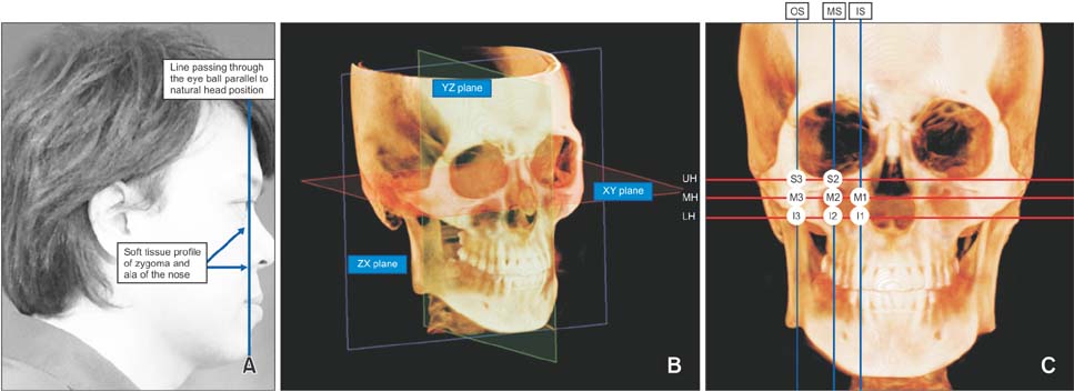
Figure 1 Inclusion criteria for the midface deficiency group. A, Soft-tissue profile with the zygoma and ala of the nose located behind the line passing through the eyeball parallel to the natural head position. B, Reference planes: XY plane, the horizontal reference plane; YZ plane, the sagittal reference plane; and ZX plane, the coronal reference plane. C, Planes parallel to each reference plane: UH, MH, LH, OS, MS, and IS. Intersecting points between the horizontal and sagittal planes were identified for middle-third facial analysis and included the S2, S3, M1, M2, M3, I1, I2, and I3.
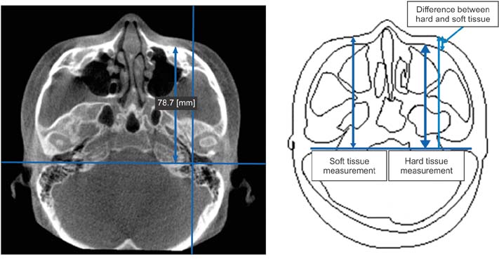
Figure 2 Linear lengths are measured from each hard-tissue (HS2, HS3, HM1, HM2, HM3, HI1, HI2, and HI3) and soft-tissue (SS2, SS3, SM1, SM2, SM3, SI1, SI2, and SI3) landmark point to the ZX plane, which paralleled the horizontal reference planes UH, MH, and LH.
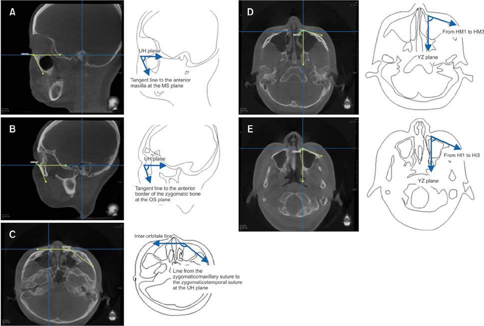
Figure 3 Angular measurements. A, Sagittal maxillary angle (SMA). B, Sagittal zygomatic angle (SZA). C, Transverse zygomatic angle (TZA). D, Middle transverse maxillary angle (MTMA). E, Inferior transverse maxillary angle (ITMA).
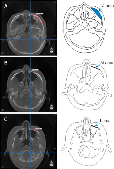
Figure 4 Surface area measurements. A, Zygomatic area (Z-area). B, Middle maxillary area (M-area). C, Inferior maxillary area (I-area).
Reference
-
1. Sabri R. Orthodontic objectives in orthognathic surgery: state of the art today. World J Orthod. 2006; 7:177–191.2. Park HC, Lee JW. Study of horizontal skeletal pattern and dental arch in skeletal Class III malocclusion patients. Korean J Orthod. 2008; 38:358–370.
Article3. Bergman RT. Cephalometric soft tissue facial analysis. Am J Orthod Dentofacial Orthop. 1999; 116:373–389.
Article4. McNeill RW, Proffit WR, White RP. Cephalometric prediction for orthodontic surgery. Angle Orthod. 1972; 42:154–164.5. Burstone CJ, James RB, Legan H, Murphy GA, Norton LA. Cephalometrics for orthognathic surgery. J Oral Surg. 1978; 36:269–277.6. Choe YK, Suhr CH. Hard and soft tissue changes after orthognathic surgery of mandibular prognathism. Korean J Orthod. 1993; 23:707–724.7. Louis PJ, Austin RB, Waite PD, Mathews CS. Soft tissue changes of the upper lip associated with maxillary advancement in obstructive sleep apnea patients. J Oral Maxillofac Surg. 2001; 59:151–156.
Article8. Stella JP, Streater MR, Epker BN, Sinn DP. Predictability of upper lip soft tissue changes with maxillary advancement. J Oral Maxillofac Surg. 1989; 47:697–703.
Article9. Betts NJ, Vig KW, Vig P, Spalding P, Fonseca RJ. Changes in the nasal and labial soft tissues after surgical repositioning of the maxilla. Int J Adult Orthodon Orthognath Surg. 1993; 8:7–23.10. Zide B, Grayson B, McCarthy JG. Cephalometric analysis for upper and lower midface surgery: Part II. Plast Reconstr Surg. 1981; 68:961–968.11. Jung JH, Kim SS, Son WS, Park SB. Soft tissue change of the midface in skeletal class III orthognathic surgery patients. Korean J Orthod. 2008; 38:83–94.
Article12. Singh GD, McNamara JA Jr, Lozanoff S. Morphometry of the midfacial complex in subjects with class III malocclusions: Procrustes, Euclidean, and cephalometric analyses. Clin Anat. 1998; 11:162–170.
Article13. Nocini PF, Boccieri A, Bertossi D. Gridplan midfacial analysis for alloplastic implants at the time of jaw surgery. Plast Reconstr Surg. 2009; 123:670–679.
Article14. Lopes PM, Moreira CR, Perrella A, Antunes JL, Cavalcanti MG. 3-D volume rendering maxillofacial analysis of angular measurements by multislice CT. Oral Surg Oral Med Oral Pathol Oral Radiol Endod. 2008; 105:224–230.
Article15. Jung YJ, Kim MJ, Baek SH. Hard and soft tissue changes after correction of mandibular prognathism and facial asymmetry by mandibular setback surgery: three-dimensional analysis using computerized tomography. Oral Surg Oral Med Oral Pathol Oral Radiol Endod. 2009; 107:763–771.e8.
Article16. Kim NK, Lee C, Kang SH, Park JW, Kim MJ, Chang YI. A three-dimensional analysis of soft and hard tissue changes after a mandibular setback surgery. Comput Methods Programs Biomed. 2006; 83:178–187.
Article17. Choi YK, Park SB, Kim YI, Son WS. Three-dimensional evaluation of midfacial asymmetry in patients with nonsyndromic unilateral cleft lip and palate by cone-beam computed tomography. Korean J Orthod. 2013; 43:113–119.
Article18. Landis JR, Koch GG. The measurement of observer agreement for categorical data. Biometrics. 1977; 33:159–174.
Article19. Chien PC, Parks ET, Eraso F, Hartsfield JK, Roberts WE, Ofner S. Comparison of reliability in anatomical landmark identification using two-dimensional digital cephalometrics and three-dimensional cone beam computed tomography in vivo. Dentomaxillofac Radiol. 2009; 38:262–273.
Article20. Hwang HS, Hwang CH, Lee KH, Kang BC. Maxillofacial 3-dimensional image analysis for the diagnosis of facial asymmetry. Am J Orthod Dentofacial Orthop. 2006; 130:779–785.
Article21. Ferrario VF, Sforza C, Serrao G, Ciusa V, Dellavia C. Growth and aging of facial soft tissues: A computerized three-dimensional mesh diagram analysis. Clin Anat. 2003; 16:420–433.
Article22. Ferrario VF, Sforza C, Schmitz JH, Santoro F. Threedimensional facial morphometric assessment of soft tissue changes after orthognathic surgery. Oral Surg Oral Med Oral Pathol Oral Radiol Endod. 1999; 88:549–556.
Article23. Soncul M, Bamber MA. Evaluation of facial soft tissue changes with optical surface scan after surgical correction of Class III deformities. J Oral Maxillofac Surg. 2004; 62:1331–1340.
Article24. Chew MT. Soft and hard tissue changes after bimaxillary surgery in Chinese Class III patients. Angle Orthod. 2005; 75:959–963.25. Harada K, Kikuchi T, Morishima S, Sato M, Ohkura K, Omura K. Changes in bite force and dentoskeletal morphology in prognathic patients after orthognathic surgery. Oral Surg Oral Med Oral Pathol Oral Radiol Endod. 2003; 95:649–654.
Article26. Cho JH, Kim EJ, Kim BC, Cho KH, Lee KH, Hwang HS. Correlations of frontal lip-line canting with craniofacial morphology and muscular activity. Am J Orthod Dentofacial Orthop. 2007; 132:278.e7–278.e14.
Article27. Kwon TG, Park HS, Ryoo HM, Lee SH. A comparison of craniofacial morphology in patients with and without facial asymmetry--a three-dimensional analysis with computed tomography. Int J Oral Maxillofac Surg. 2006; 35:43–48.
Article28. Morris DE, Moaveni Z, Lo LJ. Aesthetic facial skeletal contouring in the Asian patient. Clin Plast Surg. 2007; 34:547–556.
Article
- Full Text Links
-
- Actions
-
Cited
- CITED
-
- Close
- Share
-
- Similar articles
-
- Correction to: Hard- and soft-tissue profiles of the midface region in patients with skeletal Class III malocclusion using cone-beam computed tomography multiplanar-reconstructed image analysis
- Evaluation of facial soft tissue thickness in asymmetric mandibular deformities after orthognathic surgery
- Cone-beam computed tomography analysis of transverse dental compensation in patients with skeletal Class III malocclusion and facial asymmetry
- Soft tissue change of the midface in skeletal class III orthognathic surgery patients
- Location and shape of the mandibular lingula: Comparison of skeletal class I and class III patients using panoramic radiography and cone-beam computed tomography
