Korean J Ophthalmol.
2018 Aug;32(4):303-311. 10.3341/kjo.2017.0093.
Optical Coherence Tomography Measurement and Visual Outcome in Acute Central Retinal Artery Occlusion
- Affiliations
-
- 1Institute of Vision Research, Department of Ophthalmology, Severance Hospital, Yonsei University College of Medicine, Seoul, Korea. semekim@yuhs.ac
- 2Department of Ophthalmology, Dankook University Hospital, Dankook University College of Medicine, Cheonan, Korea.
- 3Siloam Eye Hospital, Seoul, Korea.
- KMID: 2418151
- DOI: http://doi.org/10.3341/kjo.2017.0093
Abstract
- PURPOSE
This study investigated visual acuity (VA) values and differences depending on optical coherence tomography (OCT) findings in patients with acute central retinal artery occlusion (CRAO).
METHODS
A retrospective chart review was performed on patients with acute CRAO who underwent macular and disc OCT. We evaluated changes in macular thickness and retinal nerve fiber layer (RNFL) thickness after acute CRAO onset based on OCT. We also determined the association of thickness changes with VA improvement.
RESULTS
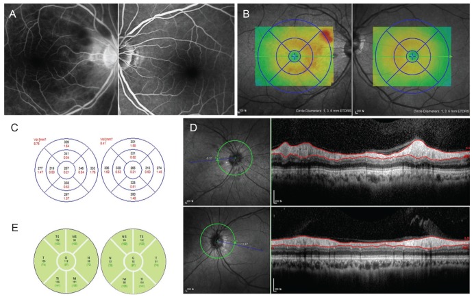
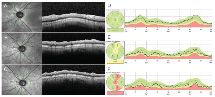
This study involved both eyes in a total of 12 patients with acute CRAO. A significant increase was observed in foveal (1 mm) thickness (p = 0.002), parafoveal (3 mm) thickness (p = 0.002), and peripapillary RNFL thickness (p = 0.005) in affected eyes with CRAO, but not in central foveal thickness (p = 0.266). A significant small difference in both eyes (affected eye - fellow eye) was shown in foveal (1 mm) and mean parafoveal (3 mm) thickness in the improved VA group (p = 0.008 and p = 0.004, respectively), but not in central foveal or peripapillary RNFL thickness (both p = 0.283).
CONCLUSIONS
Both macular and RNFL thickness increased in patients with acute CRAO. RNFL thickness decreased over time with progression of RNFL atrophy. Less macular damage caused by acute CRAO could be predicted by a small difference in macular thickness between eyes (affected eye - fellow eye). In such cases, patients had a greater chance of VA improvement.
Keyword
MeSH Terms
Figure
Reference
-
1. Sharma S, Brown GC. Retinal artery obstruction. In : Ryan SJ, editor. Retina. St. Louis: Mosby;2001. p. 1350–1367.2. Hollenhorst RW. Significance of bright plaques in the retinal arterioles. JAMA. 1961; 178:23–29. PMID: 13908419.
Article3. Rim TH, Han J, Choi YS, et al. Retinal artery occlusion and the risk of stroke development: twelve-year nationwide cohort study. Stroke. 2016; 47:376–382. PMID: 26742801.
Article4. Ahn SJ, Woo SJ, Park KH, et al. Retinal and choroidal changes and visual outcome in central retinal artery occlusion: an optical coherence tomography study. Am J Ophthalmol. 2015; 159:667–676. PMID: 25579642.
Article5. Chen H, Xia H, Qiu Z, et al. Correlation of optical intensity on optical coherence tomography and visual outcome in central retinal artery occlusion. Retina. 2016; 36:1964–1970. PMID: 26966866.
Article6. Chen SN, Hwang JF, Chen YT. Macular thickness measurements in central retinal artery occlusion by optical coherence tomography. Retina. 2011; 31:730–737. PMID: 21242861.
Article7. Yu S, Pang CE, Gong Y, et al. The spectrum of superficial and deep capillary ischemia in retinal artery occlusion. Am J Ophthalmol. 2015; 159:53–63. PMID: 25244976.
Article8. Shinoda K, Yamada K, Matsumoto CS, et al. Changes in retinal thickness are correlated with alterations of electroretinogram in eyes with central retinal artery occlusion. Graefes Arch Clin Exp Ophthalmol. 2008; 246:949–954. PMID: 18425524.
Article9. Schmidt DP, Schulte-Monting J, Schumacher M. Prognosis of central retinal artery occlusion: local intraarterial fibrinolysis versus conservative treatment. AJNR Am J Neuroradiol. 2002; 23:1301–1307. PMID: 12223369.10. Hayreh SS, Zimmerman MB. Fundus changes in central retinal artery occlusion. Retina. 2007; 27:276–289. PMID: 17460582.
Article11. Hayreh SS. Acute retinal arterial occlusive disorders. Prog Retin Eye Res. 2011; 30:359–394. PMID: 21620994.
Article12. Rumelt S, Brown GC. Update on treatment of retinal arterial occlusions. Curr Opin Ophthalmol. 2003; 14:139–141. PMID: 12777932.
Article13. Cugati S, Varma DD, Chen CS, Lee AW. Treatment options for central retinal artery occlusion. Curr Treat Options Neurol. 2013; 15:63–77. PMID: 23070637.
Article14. Ahn SJ, Kim JM, Hong JH, et al. Efficacy and safety of intra-arterial thrombolysis in central retinal artery occlusion. Invest Ophthalmol Vis Sci. 2013; 54:7746–7755. PMID: 24176901.
Article15. Hattenbach LO, Kuhli-Hattenbach C, Scharrer I, Baatz H. Intravenous thrombolysis with low-dose recombinant tissue plasminogen activator in central retinal artery occlusion. Am J Ophthalmol. 2008; 146:700–706. PMID: 18718570.
Article16. Chen CS, Lee AW, Campbell B, et al. Efficacy of intravenous tissue-type plasminogen activator in central retinal artery occlusion: report from a randomized, controlled trial. Stroke. 2011; 42:2229–2234. PMID: 21757667.17. Schumacher M, Schmidt D, Jurklies B, et al. Central retinal artery occlusion: local intra-arterial fibrinolysis versus conservative treatment, a multicenter randomized trial. Ophthalmology. 2010; 117:1367–1375. PMID: 20609991.
Article18. Schrag M, Youn T, Schindler J, et al. Intravenous fibrinolytic therapy in central retinal artery occlusion: a patient-level meta-analysis. JAMA Neurol. 2015; 72:1148–1154. PMID: 26258861.19. Ikeda F, Kishi S. Inner neural retina loss in central retinal artery occlusion. Jpn J Ophthalmol. 2010; 54:423–429. PMID: 21052904.
Article20. Klein R, Klein BE, Moss SE, Meuer SM. Retinal emboli and cardiovascular disease: the Beaver Dam Eye Study. Arch Ophthalmol. 2003; 121:1446–1451. PMID: 14557181.
- Full Text Links
- Actions
-
Cited
- CITED
-
- Close
- Share
- Similar articles
-
- Spectral-Domain Optical Coherence Tomography Findings in Acute Central Retinal Artery Occlusion
- Clinical Manifestations and Visual Prognosis of Cilioretinal Artery Sparing Central Retinal Artery Occlusion
- The Use fulness of OCT[Optical Coherence Tomography]for the Diagnosis of Central Serous Choriore tinopathy
- Patterns of Macular Edema in Patients with Branch Retinal Vein Occlusion on Optical Coherence Tomography
- A Case of Central Retinal Artery Occlusion after Intravitreal Triamcinolone Acetonide Injection for Diabetic Macular Edema in Non-Proliferative Diabetic Retinopathy


