Korean J Ophthalmol.
2018 Aug;32(4):249-256. 10.3341/kjo.2017.0118.
Clinical and Demographic Characteristics of Blepharoptosis in Korea: A 24-year Experience including 2,328 Patients
- Affiliations
-
- 1Department of Ophthalmology, Kim's Eye Hospital, Myung-Gok Eye Research Institute, Seoul, Korea. ecykim@kimeye.com
- 2Department of Ophthalmology, Kyungpook National University School of Medicine, Daegu, Korea.
- 3Department of Ophthalmology, Konyang University College of Medicine, Daejon, Korea.
- 4Lee's Eye Clinic, Seoul, Korea.
- 5Institute of Vision Research, Department of Ophthalmology, Yonsei University College of Medicine, Seoul, Korea.
- KMID: 2418144
- DOI: http://doi.org/10.3341/kjo.2017.0118
Abstract
- PURPOSE
To describe the demographics, relative incidence of subtypes, and clinical characteristics of blepharoptosis in Korean patients.
METHODS
This is a retrospective, observational case series consisting of 2,328 patients who underwent ptosis surgery from 1991 to 2014 at a tertiary referral hospital in Korea. The patients were classified according to the type of ptosis and the evaluation of clinical characteristics including levator muscle function (LF) and degree of ptosis.
RESULTS
Of the 2,328 patients, 1,815 (78%) had congenital ptosis and 513 (22%) had acquired ptosis. Simple congenital ptosis is the most common type overall (73.7%), and aponeurotic ptosis is the most common acquired type. More than three-quarters of eyes with congenital ptosis were affected in a moderate (34.4%) to severe degree (41.3%), and most of these eyes had fair (33.7%) to poor LF (60.1%). Among eyes with acquired ptosis, approximately three-quarters were affected in a mild (33.3%) to moderate degree (41.0%), with 63.3% of these eyes having good LF. The most widely used surgical technique was frontalis suspension (55.1%), followed by levator resection (29.0%) and aponeurosis repair (14.8%). At 3 years after the first surgery, 15.7% of patients with congenital ptosis and 10.4% of patients with acquired ptosis underwent reoperation.
CONCLUSIONS
Although the prevalence has decreased from previous years, the proportion of cases with congenital ptosis was higher in this study than has been shown in research conducted in the West. The majority of eyes with congenital ptosis was affected to a severe degree and had poor LF, while those with acquired ptosis were affected to a moderate degree and had good LF. More cases with acquired ptosis presented with fair to poor LF, and frontalis suspension surgery was performed more commonly compared to previous studies. The reoperation rate was higher in congenital ptosis compared to acquired ptosis.
Keyword
MeSH Terms
Figure
-
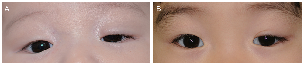
Fig. 1 Standard photographs for evaluating levator muscle function (LF) of a young patient whose LF could not be measured accurately. (A) An inert eyelid, extensive eyebrow elevation, no double eyelid crease, severe lid drooping, and chin lifting head posture suggested poor LF (left eye), (B) compared with fair LF suggested by some movement of the eyelid especially on vertical gaze, a double eyelid crease, and mild lid drooping (left eye).
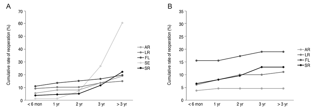
Fig. 2 The cumulative rate of reoperation in (A) congenital and (B) acquired ptosis according to surgical method. AR = aponeurosis repair; LR = levator resection; FL = autogenous fascia lata suspension; SE = Supramid Extra suspension; SR = silicone rod suspension.
Reference
-
1. Frueh BR. The mechanistic classification of ptosis. Ophthalmology. 1980; 87:1019–1021.
Article2. Fox SA. Ophthalmic plastic surgery. 5th ed. New York: Grune & Stratton;1976. p. 353–359.3. Beard C. Ptosis. 3rd ed. St. Louis: Mosby;1981. p. 39–75.4. Berke RN. Congenital ptosis; a classification of 200 cases. Arch Ophthal. 1949; 41:188–197.5. Carbajal UM. An analysis of 142 cases of ptosis. Am J Ophthalmol. 1958; 45:692–704.
Article6. Smith B, McCord CD, Baylis H. Surgical treatment of blepharoptosis. Am J Ophthalmol. 1969; 68:92–99.
Article7. Clauser L, Tieghi R, Galie M. Palpebral ptosis: clinical classification, differential diagnosis, and surgical guidelines: an overview. J Craniofac Surg. 2006; 17:246–254.8. Nerad JA. Evaluation and treatment of the patient with ptosis. In : Nerad JA, editor. Oculoplastic surgery: the requisites in ophthalmology. St. Louis: Mosby;2001. p. 157–192.9. Beard C. A new classification of blepharoptosis. Int Ophthalmol Clin. 1989; 29:214–216.
Article10. de Figueiredo AR. Blepharoptosis. Semin Ophthalmol. 2010; 25:39–51.11. Fox SA. A new ptosis classification: late spontaneous ptosis. Arch Ophthalmol. 1972; 88:590–593.12. Rathbun JE. Eyelid surgery. 1st ed. Boston: Little Brown;1990. p. 201–217.13. Lim JM, Hou JH, Singa RM, et al. Relative incidence of blepharoptosis subtypes in an oculoplastics practice at a tertiary care center. Orbit. 2013; 32:231–234.
Article14. Ahn YS, Lee TS. Clinical observation and their surgical results of 67 cases of blepharoptosis. J Korean Ophthalmol Soc. 1979; 20:283–290.15. Kim HM, Lee TS. Clinical observation and their surgical results of 127 cases of blepharoptosis. J Korean Ophthalmol Soc. 1985; 26:441–448.16. Kim SY, Chung WS. Analysis of the causes of ptosis. J Korean Ophthalmol Soc. 1995; 36:1649–1654.17. Kim IS, Choi JB, Rah SH, Lee SY. Classification of ptosis in Korea. J Korean Ophthalmol Soc. 2005; 46:1262–1269.18. Griepentrog GJ, Diehl NN, Mohney BG. Incidence and demographics of childhood ptosis. Ophthalmology. 2011; 118:1180–1183.
Article19. Lee V, Konrad H, Bunce C, et al. Aetiology and surgical treatment of childhood blepharoptosis. Br J Ophthalmol. 2002; 86:1282–1286.
Article20. Berry-Brincat A, Willshaw H. Paediatric blepharoptosis: a 10-year review. Eye (Lond). 2009; 23:1554–1559.
Article21. Finsterer J. Ptosis: causes, presentation, and management. Aesthetic Plast Surg. 2003; 27:193–204.
Article22. Kim CY, Lee SY. Distinct features in Koreans with involutional blepharoptosis. Plast Reconstr Surg. 2015; 135:1693–1699.
Article23. Park CY, Jeon SL, Woo KI, Chang HR. The frequency and aspects of ptosis in Korean old age. J Korean Ophthalmol Soc. 2007; 48:205–210.24. Pereira LS, Hwang TN, Kersten RC, et al. Levator superioris muscle function in involutional blepharoptosis. Am J Ophthalmol. 2008; 145:1095–1098.
Article25. Shields M, Putterman A. Blepharoptosis correction. Curr Opin Otolaryngol Head Neck Surg. 2003; 11:261–266.
Article26. Kim CY, Son BJ, Son J, et al. Analysis of the causes of recurrence after frontalis suspension using silicone rods for congenital ptosis. PLoS One. 2017; 12:e0171769.
Article
- Full Text Links
-
- Actions
-
Cited
- CITED
-
- Close
- Share
-
- Similar articles
-
- Congenital Blepharoptosis
- Blepharoptosis Secondary to Local Conjunctival and Tarsal Amyloidosis
- The Surgical Effect of Full-Thickness Eyelid Resection in Traumatic Blepharoptosis
- Treatment of Severe Blepharoptosis after Blow Out Fracture
- Frontalis Suspension for Blepharoptosis using Palmaris Longus Tendon
