Korean J Orthod.
2018 Jul;48(4):245-252. 10.4041/kjod.2018.48.4.245.
Effects of different primers on indirect orthodontic bonding: Shear bond strength, color change, and enamel roughness
- Affiliations
-
- 1Department of Orthodontics, School of Dentistry, Federal University of Rio de Janeiro, Rio de Janeiro, Brazil. linojima@gmail.com
- 2Military Institute of Engineering, Rio de Janeiro, Brazil.
- KMID: 2417966
- DOI: http://doi.org/10.4041/kjod.2018.48.4.245
Abstract
OBJECTIVE
We aimed to perform in-vitro evaluation to compare 1) shear bond strength (SBS), adhesive remnant index (ARI), and color change between self-etched and acid-etched primers; 2) the SBS, ARI and color change between direct and indirect bonding; and 3) the enamel roughness (ER) between 12-blade bur and aluminum oxide polisher debonding methods.
METHODS
Seventy bovine incisors were distributed in seven groups: control (no bonding), direct (DTBX), and 5 indirect bonding (ITBX, IZ350, ISONDHI, ISEP, and ITBXp). Transbond XT Primer was used in the DTBX, ITBX, and ITBXp groups, flow resin Z350 in the IZ350 group, Sondhi in the ISONDHI group, and SEP primer in the ISEP group. SBS, ARI, and ER were evaluated. The adhesive remnant was removed using a low-speed tungsten bur in all groups except the ITBXp, in which an aluminum oxide polisher was used. After coffee staining, color evaluations were performed using a spectrophotometer immediately after staining and prior to bonding.
RESULTS
ISONDHI and ISEP showed significantly lower SBS (p < 0.01). DTBX had a greater number of teeth with all the adhesive on the enamel (70%), compared with the indirect bonding groups (0-30%). The ER in the ITBX and ITBXp groups was found to be greater because of both clean-up techniques used.
CONCLUSIONS
Direct and indirect bonding have similar results and all the primers used show satisfactory adhesion strength. Use of burs and polishers increases the ER, but polishers ensure greater integrity of the initial roughness. Resin tags do not change the color of the teeth.
Keyword
MeSH Terms
Figure
-
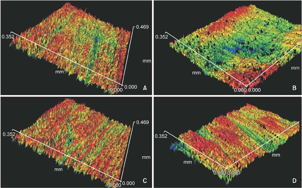
Figure 1 A, Three-dimensional (3D) profile of ITBX before bonding. B, 3D profile of ITBX after clean-up. C, 3D profile of IBTXp before bonding. D, 3D profile of ITBXp after clean-up. Red represents the peak, and blue, the valley.
Cited by 1 articles
-
Effect of orthodontic bonding with different surface treatments on color stability and translucency of full cubic stabilized zirconia after coffee thermocycling
Yasamin Babaee Hemmati, Hamid Neshandar Asli, Alireza Mahmoudi Nahavandi, Nika Safari, Mehran Falahchai
Korean J Orthod. 2023;53(3):139-149. doi: 10.4041/kjod22.144.
Reference
-
1. Newman GV. Clinical treatment with bonded plastic attachments. Am J Orthod. 1971; 60:600–610.
Article2. Sondhi A. Efficient and effective indirect bonding. Am J Orthod Dentofacial Orthop. 1999; 115:352–359.
Article3. Deahl ST, Salome N, Hatch JP, Rugh JD. Practice-based comparison of direct and indirect bonding. Am J Orthod Dentofacial Orthop. 2007; 132:738–742.
Article4. Shimizu RH, Grando KG, Shimizu IA, Andriguetto AR, Melo ACM, Witters EL. Assessment of shear bond strength of brackets bonded by direct and indirect techniques: An in vitro study. Dental Press J Orthod. 2012; 17:23.e1–23.e7.
Article5. Eliades T, Gioka C, Heim M, Eliades G, Makou M. Color stability of orthodontic adhesive resins. Angle Orthod. 2004; 74:391–393.6. Nojima LI, Araújo AS, Alves Júnior M. Indirect orthodontic bonding--a modified technique for improved efficiency and precision. Dental Press J Orthod. 2015; 20:109–117.
Article7. Artun J, Bergland S. Clinical trials with crystal growth conditioning as an alternative to acid-etch enamel pretreatment. Am J Orthod. 1984; 85:333–340.
Article8. Stober T, Gilde H, Lenz P. Color stability of highly filled composite resin materials for facings. Dent Mater. 2001; 17:87–94.
Article9. Gegauff AG, Rosenstiel SF, Langhout KJ, Johnston WM. Evaluating tooth color change from carbamide peroxide gel. J Am Dent Assoc. 1993; 124:65–72.
Article10. Oesterle LJ, Shellhart WC, Belanger GK. The use of bovine enamel in bonding studies. Am J Orthod Dentofacial Orthop. 1998; 114:514–519.
Article11. Yassen GH, Platt JA, Hara AT. Bovine teeth as substitute for human teeth in dental research: a review of literature. J Oral Sci. 2011; 53:273–282.
Article12. Reynolds IR, von Fraunhofer JA. Direct bonding in orthodontics: a comparison of attachments. Br J Orthod. 1977; 4:65–69.
Article13. Hellak A, Ebeling J, Schauseil M, Stein S, Roggendorf M, Korbmacher-Steiner H. Shear bond strength of three orthodontic bonding systems on enamel and restorative materials. Biomed Res Int. 2016; 2016:6307107.
Article14. Linn BJ, Berzins DW, Dhuru VB, Bradley TG. A comparison of bond strength between direct- and indirect-bonding methods. Angle Orthod. 2006; 76:289–294.15. Yi GK, Dunn WJ, Taloumis LJ. Shear bond strength comparison between direct and indirect bonded orthodontic brackets. Am J Orthod Dentofacial Orthop. 2003; 124:577–581.
Article16. Menini A, Cozzani M, Sfondrini MF, Scribante A, Cozzani P, Gandini P. A 15-month evaluation of bond failures of orthodontic brackets bonded with direct versus indirect bonding technique: a clinical trial. Prog Orthod. 2014; 15:70.
Article17. Daub J, Berzins DW, Linn BJ, Bradley TG. Bond strength of direct and indirect bonded brackets after thermocycling. Angle Orthod. 2006; 76:295–300.18. Polat O, Karaman AI, Buyukyilmaz T. In vitro evaluation of shear bond strengths and in vivo analysis of bond survival of indirect-bonding resins. Angle Orthod. 2004; 74:405–409.19. Klocke A, Shi J, Kahl-Nieke B, Bismayer U. Bond strength with custom base indirect bonding techniques. Angle Orthod. 2003; 73:176–180.20. Miles PG, Weyant RJ. A comparison of two indirect bonding adhesives. Angle Orthod. 2005; 75:1019–1023.21. Goracci C, Margvelashvili M, Giovannetti A, Vichi A, Ferrari M. Shear bond strength of orthodontic brackets bonded with a new self-adhering flowable resin composite. Clin Oral Investig. 2013; 17:609–617.
Article22. Diedrich P. Enamel alterations from bracket bonding and debonding: a study with the scanning electron microscope. Am J Orthod. 1981; 79:500–522.
Article23. Kim SS, Park WK, Son WS, Ahn HS, Ro JH, Kim YD. Enamel surface evaluation after removal of orthodontic composite remnants by intraoral sandblasting: a 3-dimensional surface profilometry study. Am J Orthod Dentofacial Orthop. 2007; 132:71–76.
Article24. Sigilião LC, Marquezan M, Elias CN, Ruellas AC, Sant'Anna EF. Efficiency of different protocols for enamel clean-up after bracket debonding: an in vitro study. Dental Press J Orthod. 2015; 20:78–85.
Article25. Janiszewska-Olszowska J, Tomkowski R, Tandecka K, Stepien P, Szatkiewicz T, Sporniak-Tutak K, et al. Effect of orthodontic debonding and residual adhesive removal on 3D enamel microroughness. PeerJ. 2016; 4:e2558.26. Karan S, Kircelli BH, Tasdelen B. Enamel surface roughness after debonding. Angle Orthod. 2010; 80:1081–1088.
Article27. Zachrisson BU, Arthun J. Enamel surface appearance after various debonding techniques. Am J Orthod. 1979; 75:121–127.
Article28. Campbell PM. Enamel surfaces after orthodontic bracket debonding. Angle Orthod. 1995; 65:103–110.29. Yannikakis SA, Zissis AJ, Polyzois GL, Caroni C. Color stability of provisional resin restorative materials. J Prosthet Dent. 1998; 80:533–539.
Article
- Full Text Links
-
- Actions
-
Cited
- CITED
-
- Close
- Share
-
- Similar articles
-
- The shear bond strength of two adhesives bonded to composite resin and glass ionomer cement restorations
- Effects of phosphoric acid concentration on depth of etch and shear bond strength of orthodontic brackets to bovine enamel
- Shear bond strength of orthodontic bonding resins to porcelain; an in vitro study
- The effects of fluoride releasing orthodontic sealant on the shear bond strength of light-and chemical-cured orthodontic resins
- The effects of crystal growth on shear bond strength of orthodontic bracket adhesives to enamel surface
