Allergy Asthma Respir Dis.
2018 Jul;6(4):229-233. 10.4168/aard.2018.6.4.229.
Ecthyma gangrenosum and agranulocytosis in a previously healthy 12-month-old girl: Report of 1 case with a literature review
- Affiliations
-
- 1Department of Pediatrics, Soonchunhyang Unversity College of Medicine, Cheonan, Korea. pjstable@schmc.ac.kr
- 2Department of Dermatology, Soonchunhyang Unversity College of Medicine, Cheonan, Korea.
- 3Department of Orthopedic Surgery, Soonchunhyang Unversity College of Medicine, Cheonan, Korea.
- 4Department of Clinical Laboratory Medicine, Soonchunhyang Unversity College of Medicine, Cheonan, Korea.
- 5Department of Emergency Medicine, Soonchunhyang Unversity College of Medicine, Cheonan, Korea.
- KMID: 2417851
- DOI: http://doi.org/10.4168/aard.2018.6.4.229
Abstract
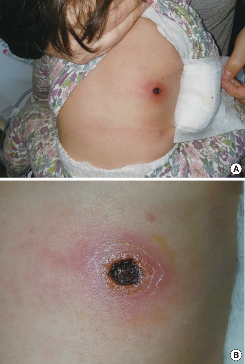
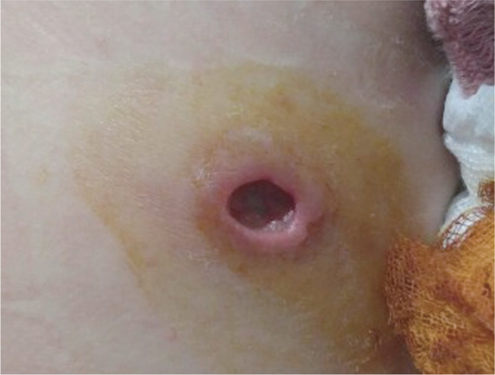
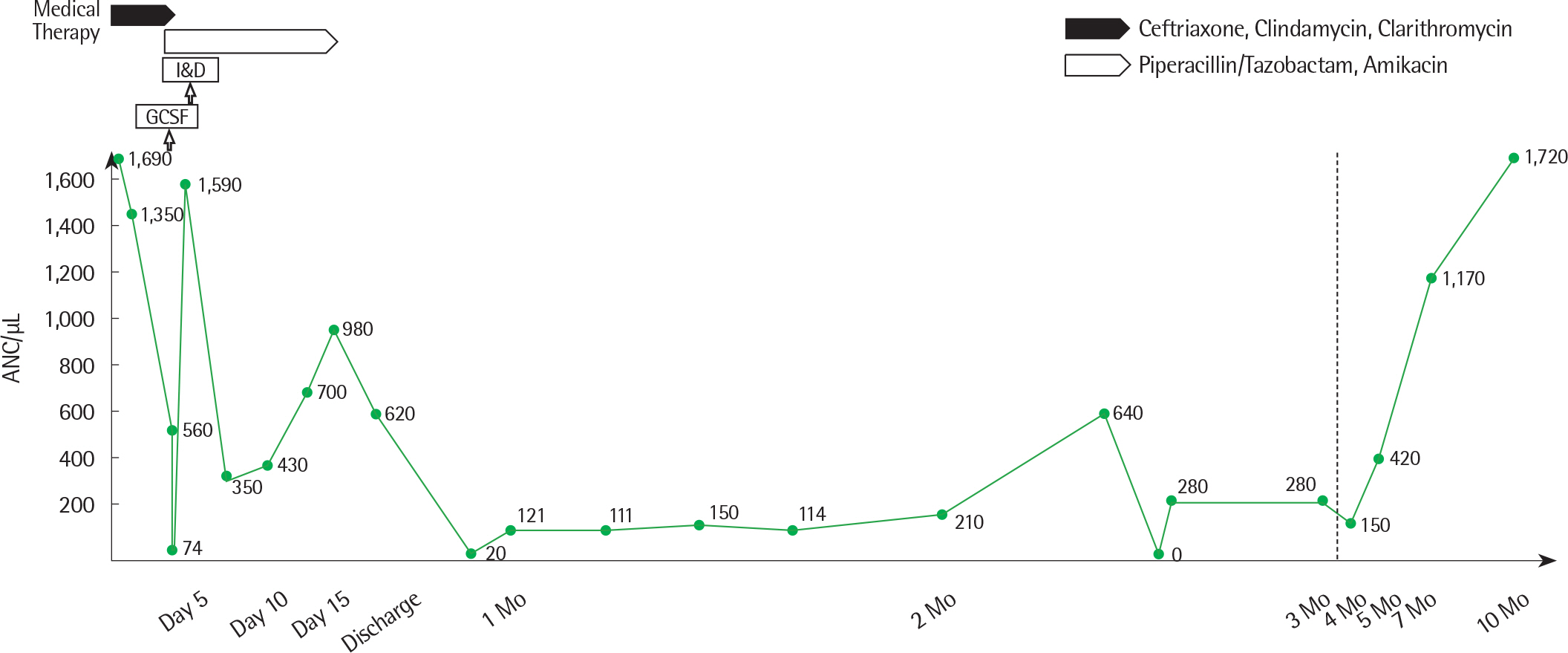
- Ecthyma gangrenosum (EG) is a rare skin manifestation which starts with a maculopapular eruption and followed by a necrotic ulcer covered with black eschar. EG usually occurs in immunosuppressed patients with Pseudomonas aeruginosa sepsis. We present a previously healthy 12-month-old girl with EG by P. aeruginosa and agranulocytosis due to influenza A and then rhinovirus infection, without bacteremia. It is important for allergists to culture wound and differentiate EG from other skin disorders including Tsutsugamushi disease and initiate appropriate empiric antipseudomonal antibiotic treatment, and to evaluate for possible immunodeficiency, even in a healthy child.
MeSH Terms
Figure
Reference
-
1. Vaiman M, Lazarovitch T, Heller L, Lotan G. Ecthyma gangrenosum and ecthyma-like lesions: review article. Eur J Clin Microbiol Infect Dis. 2015; 34:633–9.
Article2. Lee MK, Yoo SY, Hwang PH. Ecthyma gangrenosum caused by Klebsiella pneumoniae in immunocompromised patient associated with severe aplastic anemia. Clin Pediatr Hematol Oncol. 2013; 20:59–61.3. Cohen N, Capua T, Bilavsky E, Dias-Polak H, Levin D, Grisaru-Soen G. Ecthyma gangrenosum skin lesions in previously healthy children. Acta Paediatr. 2015; 104:e134–8.
Article4. Murray TS, Baltimore RS. Pseudomonas, burkholderia, and stenotroph-omonas. Kliegman R, Stanton B, Behrman R, Geme JS, Schor N, editors. Nelson textbook of pediatrics. 20th ed.Philadelphia (PA): Elsevier;2016. p. 1412–5.5. Koo SH, Lee JH, Shin H, Lee JI. Ecthyma gangrenosum in a previously healthy infant. Arch Plast Surg. 2012; 39:673–5.
Article6. Seo JY, Kim SY, Han MY, Lee KH. A case of ecthyma gangrenosum associated with liver abscess and renal abscess. Korean J Pediatr Infect Dis. 2002; 9:104–9.
Article7. Juern AM, Drolet BA. Ecthyma. Kliegman R, Stanton B, Behrman R, Geme JS, Schor N, editors. Nelson textbook of pediatrics. 20th ed.Philadelphia (PA): Elsevier;2016. p. 3207–8.8. Vaiman M, Lasarovitch T, Heller L, Lotan G. Ecthyma gangrenosum versus ecthyma-like lesions: should we separate these conditions? Acta Der-matovenerol Alp Pannonica Adriat. 2015; 24:69–72.
Article9. Bozkurt I, Yuksel EP, Sunbul M. Ecthyma gangrenosum in a previously healthy patient. Indian Dermatol Online J. 2015; 6:336–8.
Article10. Pacha O, Hebert AA. Ecthyma gangrenosum and neutropenia in a previously healthy child. Pediatr Dermatol. 2013; 30:e283–4.
Article11. Sharon N, Talnir R, Lavid O, Rubinstein U, Niven M, First Y, et al. Tran-sient lymphopenia and neutropenia: pediatric influenza A/H1N1 infection in a primary hospital in Israel. Isr Med Assoc J. 2011; 13:408–12.12. Abraham TY, Dat N, John NG. Rhinovirus infection among patients with hematologic malignancy at a cancer center. Infect Dis Clin Pract. 2016; 24:29–30.13. Celkan T, Koç BŞ. Approach to the patient with neutropenia in childhood. Turk Pediatri Ars. 2015; 50:136–44.
Article14. Lanzkowsky P. Disorders of white blood cell. Lanzkowsky P, editor. Manual of pediatric hematology and oncology. 5th ed.Philadelphia (PA): Elsevier;2011. p. 275–95.15. Biscaye S, Demonchy D, Afanetti M, Dupont A, Haas H, Tran A. Ecthyma gangrenosum, a skin manifestation of Pseudomonas aeruginosa sepsis in a previously healthy child: a case report. Medicine (Baltimore). 2017; 96:e5507.16. Fang LC, Peng CC, Chi H, Lee KS, Chiu NC. Pseudomonas aeruginosa sepsis with ecthyma gangrenosum and pseudomembranous pharyngo-laryngitis in a 5-month-old boy. J Microbiol Immunol Infect. 2014; 47:158–61.
Article
- Full Text Links
- Actions
-
Cited
- CITED
-
- Close
- Share
- Similar articles
-
- A Case of Ecthyma Gangrenosum Associated with Liver Abscess
- A Case of Ecthyma Gangrenosum Associated with Liver Abscess and Renal Abscess
- Ecthyma Gangrenosum in a Previously Healthy Adolescent
- A Case of Ecthyma Gangrenosum Associated with Liver Abscess and Renal Abscess
- Ecthyma gangrenosum associated with aplastic anemia


