Hepatocellular Carcinoma Arising in a Huge Hepatocellular Adenoma with Bone Marrow Metaplasia
- Affiliations
-
- 1Department of Pathology, Asan Medical Center, University of Ulsan College of Medicine, Seoul, Korea. Jihunkim@amc.seoul.kr
- 2Department of Physiology, Asan Medical Center, University of Ulsan College of Medicine, Seoul, Korea.
- 3Asan Liver Center, Asan Medical Center, University of Ulsan College of Medicine, Seoul, Korea.
- 4Department of Surgery, Asan Medical Center, University of Ulsan College of Medicine, Seoul, Korea.
- 5Department of Radiology, Asan Medical Center, University of Ulsan College of Medicine, Seoul, Korea.
- KMID: 2417806
- DOI: http://doi.org/10.4132/jptm.2017.11.12
Abstract
- Hepatocellular adenoma (HCA) is the most common type of benign liver tumor, and its major complication is malignant transformation to hepatocellular carcinoma (HCC). Here, we report a case of HCC arising in HCA with bone marrow metaplasia in a 24-year-old Korean woman who presented with abdominal discomfort. A huge liver mass was found on abdominal ultrasonography. She underwent surgical hepatic resection, and the resected specimen was entirely involved by a 20-cm-sized tumor. Histological review revealed a well differentiated HCC arising from inflammatory HCA with β-catenin nuclear positivity and bone marrow metaplasia that contained hematopoietic cells. This case was unique because malignant transformation, inflammatory type HCA, β-catenin nuclear staining, and bone marrow metaplasia were simultaneously observed. Additionally, it should be noted that a large HCA with β-catenin activation can undergo malignant transformation and should be surgically resected in a timely manner.
MeSH Terms
Figure
-

Fig. 1. Imaging and gross pathological findings. (A) Dynamic liver and pelvis computed tomography shows an enlarged liver with a huge, well-defined, hypervascular mass in segments 4, 5, and 8 of the liver during arterial phase. (B) The surface of a slice from a surgically resected specimen reveals a heterogeneously yellow-brown color and multinodular growth with partly cystic degeneration that contained hemorrahge and necrosis.

Fig. 2. Microscopic and immunohistochemical findings of hepatocellular adenoma (HCA), inflammatory type with β-catenin activation (A–F) and hepatocellular carcinoma (HCC) (G–L). (A) Dilated sinusoids and intratumoral inflammation in the portal tract-like areas are observed in the parenchyma. (B) Large aberrant arteries with infiltration of variable inflammatory cells including lymphocytes, neutrophils, and eosinophils are occasionally observed. Tumor cells show diffuse cytoplasmic expression of serum amyloid A (C), C-reactive protein (D), and glutamine synthetase (E). (F) Nuclear β-catenin expression (arrows) is observed in several tumor cells. (G) Nuclear atypia and thick trabeculae are observed in the HCC component. (H) Heat shock protein-70 is strongly and diffusely positive. (I) Glypican 3 is partially positive. (J) Loss of reticulin is observed in the HCC component, whereas reticulin staining is preserved in the HCA (inset). (K) CD34 expression on sinusoidal cells is strong and diffuse in HCC but is patchy in HCA (inset). (L) The Ki-67 labeling index of the HCC area is significantly increased compared with that of HCA (inset).
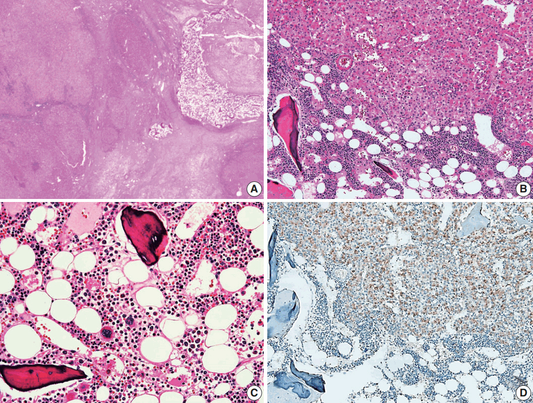
Fig. 3. Histological findings of bone marrow metaplasia. (A) Bone marrow metaplasia is observed in the inner hepatocellular adenoma. (B, C) Bone marrow metaplasia is characterized by mature lamellar bone that formed trabeculae intermingled with fat tissue containing erythroblasts, myeloblasts, and megakaryocytes. (D) Tumor cells near the bone marrow metaplasia shows positivity for epithelial cell adhesion molecule immunohistochemical staining.
Reference
-
1. Chang CY, Hernandez-Prera JC, Roayaie S, Schwartz M, Thung SN. Changing epidemiology of hepatocellular adenoma in the United States: review of the literature. Int J Hepatol. 2013; 2013:604860.
Article2. Bioulac-Sage P, Sempoux C, Balabaud C. Hepatocellular adenoma: classification, variants and clinical relevance. Semin Diagn Pathol. 2017; 34:112–25.
Article3. Bioulac-Sage P, Balabaud C, Zucman-Rossi J. Subtype classification of hepatocellular adenoma. Dig Surg. 2010; 27:39–45.
Article4. Margolskee E, Bao F, de Gonzalez AK, et al. Hepatocellular adenoma classification: a comparative evaluation of immunohistochemistry and targeted mutational analysis. Diagn Pathol. 2016; 11:27.
Article5. Chu HH, Moon WS. β-catenin activated hepatocellular adenoma. Clin Mol Hepatol. 2013; 19:185–9.
Article6. Stoot JH, Coelen RJ, De Jong MC, Dejong CH. Malignant transformation of hepatocellular adenomas into hepatocellular carcinomas: a systematic review including more than 1600 adenoma cases. HPB (Oxford). 2010; 12:509–22.
Article7. Copin P, Ronot M, Vilgrain V. Hepatocellular carcinoma with osseous metaplasia and bone marrow elements. Clin Gastroenterol Hepatol. 2015; 13:e26–7.
Article8. Iguchi T, Yamagata M, Sonoda T, et al. Malignant transformation of hepatocellular adenoma with bone marrow metaplasia arising in glycogen storage disease type I: a case report. Mol Clin Oncol. 2016; 5:599–603.
Article9. Moriura S, Kuroda M, Kimura A, et al. Case report: hepatic adenoma with bone marrow metaplasia in a patient with glycogen storage disease type 1a. J Gastroenterol Hepatol. 1996; 11:556–9.
Article10. Ramacciato G, Nigri GR, Aurello P, et al. Giant hepatic adenoma with bone marrow metaplasia not associated with oral contraceptive intake. World J Surg Oncol. 2006; 4:58.
Article11. Vaithianathan R, Selvambigai G, Jayaganesh P. Spontaneous hepatocellular adenoma in paediatric age group: case report. J Clin Diagn Res. 2013; 7:2962–3.12. Velazquez I, Alter BP. Androgens and liver tumors: Fanconi’s anemia and non-Fanconi's conditions. Am J Hematol. 2004; 77:257–67.
Article13. Stueck AE, Qu Z, Huang MA, Campreciós G, Ferrell LD, Thung SN. Hepatocellular carcinoma arising in an HNF-1alpha-mutated adenoma in a 23-year-old woman with maturity-onset diabetes of the young: a case report. Semin Liver Dis. 2015; 35:444–9.14. Hirata E, Shimizu S, Umeda S, et al. Hepatocyte nuclear factor 1alpha-inactivated hepatocellular adenomatosis in a patient with maturity-onset diabetes of the young type 3: case report and literature review. Nihon Shokakibyo Gakkai Zasshi. 2015; 112:1696–704.15. Dokmak S, Paradis V, Vilgrain V, et al. A single-center surgical experience of 122 patients with single and multiple hepatocellular adenomas. Gastroenterology. 2009; 137:1698–705.
Article16. Gorayski P, Thompson CH, Subhash HS, Thomas AC. Hepatocellular carcinoma associated with recreational anabolic steroid use. Br J Sports Med. 2008; 42:74–5.
Article17. Zucman-Rossi J, Jeannot E, Nhieu JT, et al. Genotype-phenotype correlation in hepatocellular adenoma: new classification and relationship with HCC. Hepatology. 2006; 43:515–24.
Article18. Franco LM, Krishnamurthy V, Bali D, et al. Hepatocellular carcinoma in glycogen storage disease type Ia: a case series. J Inherit Metab Dis. 2005; 28:153–62.
Article19. Terkivatan T, de Wilt JH, de Man RA, et al. Indications and longterm outcome of treatment for benign hepatic tumors: a critical appraisal. Arch Surg. 2001; 136:1033–8.20. Herzog EL, Chai L, Krause DS. Plasticity of marrow-derived stem cells. Blood. 2003; 102:3483–93.
Article
- Full Text Links
-
- Actions
-
Cited
- CITED
-
- Close
- Share
-
- Similar articles
-
- Hepatocellular Carcinoma Arising in Hepatocellular Adenoma
- Hepatocellular Carcinoma Arising from Hepatocellular Adenoma in an Elderly Male Patient
- Atypical Nodule Arising in a Hepatocellular Adenoma
- Gelatinous transformation of the bone marrow in hepatocellular carcinoma
- A Case of a Patient Presenting with Upper Gastrointestinal Bleeding Due to Direct Stomach Invasion by Hepatocellular Carcinoma
