Isolation and characterization of Korean porcine deltacoronavirus strain KNU16-07
- Affiliations
-
- 1Animal Virology Laboratory, School of Life Sciences, BK21 Plus KNU Creative BioResearch Group, Kyungpook National University, Daegu 41566, Korea. changhee@knu.ac.kr
- 2Animal Disease Diagnostic Division, Animal and Plant Quarantine Agency, Gimcheon 39660, Korea. naturelkk@korea.kr
- 3College of Veterinary Medicine, Kyungpook National University, Daegu 41566, Korea.
- 4College of Veterinary Medicine, Jeju National University, Jeju 63243, Korea.
- KMID: 2417575
- DOI: http://doi.org/10.4142/jvs.2018.19.4.577
Abstract
- Porcine deltacoronavirus (PDCoV) has emerged in several pig-raising countries and has been a causative pathogen associated with diarrheal diseases in South Korea since 2014. In the present study, we were able to isolate and cultivate a Korean PDCoV strain (KNU16-07) in cell culture and investigate its pathogenicity. PDCoV-inoculated piglets showed watery diarrhea accompanied by acute enteritis in the natural host. Sequencing analysis demonstrated the genetic stability of KNU16-07 for at least thirty serial passages.
Figure
-
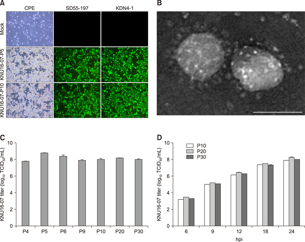
Fig. 1 Cytopathology and growth properties of porcine deltacoronavirus (PDCoV) isolate KNU16-07 in infected swine testicle (ST) cells. (A) Cytopathic effect (CPE) formation in ST cells infected with PDCoV KNU16-07-P5 or -P10. PDCoV-specific CPEs were observed daily and were photographed at 24 h post-infection (hpi) using an inverted microscope. The infected cells were subjected to immunofluorescence assay using the indicated N-specific monoclonal antibodies and examined using a fluorescence microscope. (B) Electron microscopy images of PDCoV KNU16-07. Purified virions were negatively stained with 2% phosphotungstic acid and viewed by using transmission electron microscope. (C) Virus titers at selected passages. ST cells were infected with PDCoV KNU16-07 harvested at the indicated passage numbers. At 24 hpi, the virus supernatants were collected and virus titers were determined. (D) One-step growth kinetics for the KNU16-07 strain. At the indicated times post-infection, culture supernatants were harvested and virus titers determined. Results are expressed as mean values from triplicate wells; error bars represent SD. TCID50, 50% tissue culture infective dose; P, passage. 200× (A), 100,000× (B). Scale bar = 100 nm (B).

Fig. 2 Macroscopic and microscopic examinations of intestines of piglets inoculated with the Korean porcine deltacoronavirus (PDCoV) strain KNU16-07. (A) Small intestine of an inoculated pig at 3 days post-inoculation (dpi) showing thin and transparent intestinal walls (arrows) and an extended stomach filled with curdled milk. (B and C) H&E stained jejunum of a KNU16-07-inoculated pig at 3 dpi showing acute diffuse and severe atrophic enteritis with moderate vacuolation (arrows) of enterocytes lining the epithelium of atrophied villi. (D) Detection of PDCoV antigen by immunohistochemistry analysis of jejunum tissue sections from a virus-inoculated piglet at 3 dpi. The PDCoV antigen signals (arrows) appear red and were detected in villous epithelial cells. 100× (B and D), 200× (C).
Reference
-
1. Chen Q, Gauger P, Stafne M, Thomas J, Arruda P, Burrough E, Madson D, Brodie J, Magstadt D, Derscheid R, Welch M, Zhang J. Pathogenicity and pathogenesis of a United States porcine deltacoronavirus cell culture isolate in 5-day-old neonatal piglets. Virology. 2015; 482:51–59.
Article2. Dong N, Fang L, Yang H, Liu H, Du T, Fang P, Wang D, Chen H, Xiao S. Isolation, genomic characterization, and pathogenicity of a Chinese porcine deltacoronavirus strain CHN-HN-2014. Vet Microbiol. 2016; 196:98–106.
Article3. Hu H, Jung K, Vlasova AN, Chepngeno J, Lu Z, Wang Q, Saif LJ. Isolation and characterization of porcine deltacoronavirus from pigs with diarrhea in the United States. J Clin Microbiol. 2015; 53:1537–1548.
Article4. Jang G, Lee KK, Kim SH, Lee C. Prevalence, complete genome sequencing and phylogenetic analysis of porcine deltacoronavirus in South Korea, 2014-2016. Transbound Emerg Dis. 2017; 64:1364–1370.
Article5. Jung K, Hu H, Eyerly B, Lu Z, Chepngeno J, Saif LJ. Pathogenicity of 2 porcine deltacoronavirus strains in gnotobiotic pigs. Emerg Infect Dis. 2015; 21:650–654.
Article6. Lee S, Kim Y, Lee C. Isolation and characterization of a Korean porcine epidemic diarrhea virus strain KNU-141112. Virus Res. 2015; 208:215–224.
Article7. Lee S, Lee C. Complete genome characterization of Korean porcine deltacoronavirus strain KOR/KNU14-04/2014. Genome Announc. 2014; 2.
Article8. Lee S, Lee C. Functional characterization and proteomic analysis of the nucleocapsid protein of porcine deltacoronavirus. Virus Res. 2015; 208:136–145.
Article9. Lee YN, Lee C. Complete genome sequence of a novel porcine parainfluenza virus 5 isolate in Korea. Arch Virol. 2013; 158:1765–1772.
Article10. Marthaler D, Jiang Y, Collins J, Rossow K. Complete genome sequence of strain SDCV/USA/Illinois121/2014, a porcine deltacoronavirus from the United States. Genome Announc. 2014; 2:e00218-14.
Article11. Marthaler D, Raymond L, Jiang Y, Collins J, Rossow K, Rovira A. Rapid detection, complete genome sequencing, and phylogenetic analysis of porcine deltacoronavirus. Emerg Infect Dis. 2014; 20:1347–1350.
Article12. Pan X, Kong N, Shan T, Zheng H, Tong W, Yang S, Li G, Zhou E, Tong G. Monoclonal antibody to N protein of porcine epidemic diarrhea virus. Monoclon Antib Immunodiagn Immunother. 2015; 34:51–54.
Article13. Saeng-Chuto K, Lorsirigool A, Temeeyasen G, Vui DT, Stott CJ, Madapong A, Tripipat T, Wegner M, Intrakamhaeng M, Chongcharoen W, Tantituvanont A, Kaewprommal P, Piriyapongsa J, Nilubol D. Different lineage of porcine deltacoronavirus in Thailand, Vietnam and Lao PDR in 2015. Transbound Emerg Dis. 2017; 64:3–10.
Article14. Song D, Zhou X, Peng Q, Chen Y, Zhang F, Huang T, Zhang T, Li A, Huang D, Wu Q, He H, Tang Y. Newly emerged porcine deltacoronavirus associated with diarrhoea in swine in China: identification, prevalence and full-length genome sequence analysis. Transbound Emerg Dis. 2015; 62:575–580.
Article
- Full Text Links
-
- Actions
-
Cited
- CITED
-
- Close
- Share
-
- Similar articles
-
- Assessment of hemagglutination activity of porcine deltacoronavirus
- Discovery of Two Chrysosporium Species with Keratinolytic Activity from Field Soil in Korea
- Isolation, characterization and neutralizing activity of porcine epidemic diarrhea viruses from Vietnam
- Isolation and characterization of a new porcine epidemic diarrhea virus variant that occurred in Korea in 2014
- Porcine epidemic diarrhea viruses from Vietnam: isolation, characterization, and neutralizing activity
