Non-invasive quantification of hepatic fat content in healthy dogs by using proton magnetic resonance spectroscopy and dual gradient echo magnetic resonance imaging
- Affiliations
-
- 1Clinic of Diagnostic Imaging, Vetsuisse Faculty, University of Zurich, 8057 Zurich, Switzerland. fdelchicca@vetclinics.uzh.ch
- 2Graduate School for Cellular and Biomedical Sciences, University of Bern, 3012 Bern, Switzerland.
- 3Section of Anesthesiology, Equine Department, Vetsuisse Faculty, University of Zurich, 8057 Zurich, Switzerland.
- 4Institute of Biomedical Engineering, University of Zurich, 8057 Zurich, Switzerland.
- 5Swiss Federal Institute of Technology (ETH Zurich), 8092 Zurich, Switzerland.
- 6Institute of Veterinary Pathology, Vetsuisse Faculty, University of Zurich, 8057 Zurich, Switzerland.
- 7Institute of Animal Nutrition, Vetsuisse Faculty, University of Zurich, 8057 Zurich, Switzerland.
- KMID: 2417574
- DOI: http://doi.org/10.4142/jvs.2018.19.4.570
Abstract
- The objective of the present study was to describe two non-invasive methods for fat quantification in normal canine liver by using magnetic resonance imaging (MRI) and spectroscopy. Eleven adult beagle dogs were anesthetized and underwent magnetic resonance examination of the cranial abdomen by performing morphologic, modified Dixon (mDixon) dual gradient echo sequence, and proton magnetic resonance spectroscopy (¹H MRS) imaging. In addition, ultrasonographic liver examination was performed, fine-needle liver aspirates and liver biopsies were obtained, and hepatic triglyceride content was assayed. Ultrasonographic, cytologic, and histologic examination results were unremarkable in all cases. The median hepatic fat fraction calculated was 2.1% (range, 1.3%-5.5%) using mDixon, 0.3% (range, 0.1%-1.0%) using ¹H MRS, and 1.6% (range 1.0%-2.5%) based on triglyceride content. The hepatic fat fractions calculated using mDixon and ¹H MRS imaging were highly correlated to that based on triglyceride content. A weak correlation between mDixon and ¹H MRS imaging was detected. The results show that hepatic fat content can be estimated using non-invasive techniques (mDixon or ¹H MRS) in healthy dogs. Further studies are warranted to evaluate the use of these techniques in dogs with varying hepatic fat content and different hepatic disorders.
Keyword
MeSH Terms
Figure
-
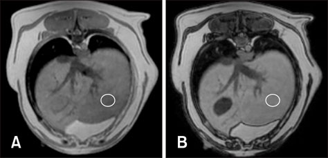
Fig. 1 Representative example of the appearance of a normal liver in the in-phase (A) and opposed-phase (B) imaging from dual gradient echo magnetic resonance imaging sequences. A regions of interest drawn in the left caudal parenchyma is shown (circles). The signal intensity is similar on both images and the mean calculated hepatic fat fraction was 1.1%.
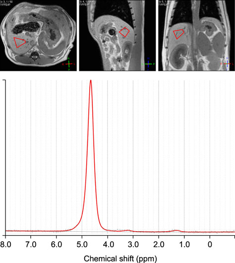
Fig. 2 Representative proton magnetic resonance spectroscopy spectrum of the dog from Fig. 1 showing the corresponding transverse (left image), sagittal (middle image) and dorsal (right image) localizing scans and the voxel placement (red polygon). On the graph, the x-axis indicates the chemical shift in parts per million (ppm). The highest peak, at the 4.6 ppm position, corresponds to the water peak. The smaller peak at the 1.2 ppm, corresponds to the lipid peak. The calculated hepatic fat fraction was 0.63%.
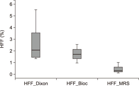
Fig. 3 Box plots of hepatic fat fraction (HFF) measured by using dual gradient echo magnetic resonance imaging (modified Dixon), proton magnetic resonance spectroscopy (MRS), and biochemical analysis (Bioc).
Reference
-
1. Borra RJ, Salo S, Dean K, Lautamäki R, Nuutila P, Komu M, Parkkola R. Nonalcoholic fatty liver disease: rapid evaluation of liver fat content with in-phase and out-of-phase MR imaging. Radiology. 2009; 250:130–136.
Article2. Chandarana H, Taouli B. Diffusion and perfusion imaging of the liver. Eur J Radiol. 2010; 76:348–358.
Article3. Clark MH, Larsen R, Lu W, Hoenig M. Investigation of 1H MRS for quantification of hepatic triglyceride in lean and obese cats. Res Vet Sci. 2013; 95:678–680.
Article4. Cullen JM, van den Ingh TSGAM, Van Winkle T, Charles JA, Desmet VJ. Morphological classification of parenchymal disorders of the canine and feline liver: 1. Normal histology, reversible hepatocytic injury and hepatic amyloidosis. WSAVA Liver Standardization Group. In : Rothuizen J, Bunch SE, Charles JA, Cullen JM, Desmet VJ, Szatmári V, Twedt DC, van den Ingh TSGAM, Van Winkle T, Washabau RJ, editors. WSAVA Standards for Clinical and Histological Diagnosis of Canine and Feline Liver Disease. Philadelphia: Elsevier;2006. p. 77–83.5. Drost WT, Henry GA, Meinkoth JH, Woods JP, Lehenbauer TW. Quantification of hepatic and renal cortical echogenicity in clinically normal cats. Am J Vet Res. 2000; 61:1016–1020.
Article6. Feeney DA, Anderson KL, Ziegler LE, Jessen CR, Daubs BM, Hardy RM. Statistical relevance of ultrasonographic criteria in the assessment of diffuse liver disease in dogs and cats. Am J Vet Res. 2008; 69:212–221.
Article7. Feeney DA, Sharkey LC, Steward SM, Bahr KL, Henson MS, Ito D, O'Brien TD, Jessen CR, Husbands BD, Borgatti A, Modiano JF. Parenchymal signal intensity in 3-T body MRI of dogs with hematopoietic neoplasia. Comp Med. 2013; 63:174–182.8. Gaschen L. Update on hepatobiliary imaging. Vet Clin North Am Small Anim Pract. 2009; 39:439–467.
Article9. Guillot M, Danjou MA, Alexander K, Bédard C, Desnoyers M, Beauregard G, Del Castillo JR. Can sonographic findings predict the results of liver aspirates in dogs with suspected liver disease? Vet Radiol Ultrasound. 2009; 50:513–518.
Article10. Guiu B, Loffroy R, Petit JM, Aho S, Ben Salem D, Masson D, Hillon P, Cercueil JP, Krause D. Mapping of liver fat with triple-echo gradient echo imaging: validation against 3.0-T proton MR spectroscopy. Eur Radiol. 2009; 19:1786–1793.
Article11. Hussain HK, Chenevert TL, Londy FJ, Gulani V, Swanson SD, McKenna BJ, Appelman HD, Adusumilli S, Greenson JK, Conjeevaram HS. Hepatic fat fraction: MR imaging for quantitative measurement and display--early experience. Radiology. 2005; 237:1048–1055.
Article12. Kemp SD, Panciera DL, Larson MM, Saunders GK, Werre SR. A comparison of hepatic sonographic features and histopathologic diagnosis in canine liver disease: 138 cases. J Vet Intern Med. 2013; 27:806–813.
Article13. Kemp SD, Zimmerman KL, Panciera DL, Monroe WE, Leib MS. Histopathologic variation between liver lobes in dogs. J Vet Intern Med. 2015; 29:58–62.
Article14. Kühn JP, Evert M, Friedrich N, Kannengiesser S, Mayerle J, Thiel R, Lerch MM, Dombrowski F, Mensel B, Hosten N, Puls R. Noninvasive quantification of hepatic fat content using three-echo dixon magnetic resonance imaging with correction for T2* relaxation effects. Invest Radiol. 2011; 46:783–789.
Article15. Lee SS, Park SH, Kim HJ, Kim SY, Kim MY, Kim DY, Suh DJ, Kim KM, Bae MH, Lee JY, Lee SG, Yu ES. Non-invasive assessment of hepatic steatosis: prospective comparison of the accuracy of imaging examinations. J Hepatol. 2010; 52:579–585.
Article16. Marks AL, Hecht S, Stokes JE, Conklin GA, Deanna KH. Effects of gadoxetate disodium (Eovist®) contrast on magnetic resonance imaging characteristics of the liver in clinically healthy dogs. Vet Radiol Ultrasound. 2014; 55:286–291.
Article17. Marsman HA, van Werven JR, Nederveen AJ, Ten Kate FJ, Heger M, Stoker J, van Gulik TM. Noninvasive quantification of hepatic steatosis in rats using 3.0 T 1H-magnetic resonance spectroscopy. J Magn Reson Imaging. 2010; 32:148–154.
Article18. Mazhar SM, Shiehmorteza M, Sirlin CB. Noninvasive assessment of hepatic steatosis. Clin Gastroenterol Hepatol. 2009; 7:135–140.
Article19. Mennesson N, Dumortier J, Hervieu V, Milot L, Guillaud O, Scoazec JY, Pilleul F. Liver steatosis quantification using magnetic resonance imaging: a prospective comparative study with liver biopsy. J Comput Assist Tomogr. 2009; 33:672–677.20. Mitchell DG, Kim I, Chang TS, Vinitski S, Consigny PM, Saponaro SA, Ehrlich SM, Rifkin MD, Rubin R. Fatty liver. Chemical shift phase-difference and suppression magnetic resonance imaging techniques in animals, phantoms, and humans. Invest Radiol. 1991; 26:1041–1052.21. Nakamura M, Chen HM, Momoi Y, Iwasaki T. Clinical application of computed tomography for the diagnosis of feline hepatic lipidosis. J Vet Med Sci. 2005; 67:1163–1165.
Article22. Perman WH, Balci NC, Akduman I. Review of magnetic resonance spectroscopy in the liver and the pancreas. Top Magn Reson Imaging. 2009; 20:89–97.
Article23. Provencher SW. Estimation of metabolite concentrations from localized in vivo proton NMR spectra. Magn Reson Med. 1993; 30:672–679.
Article24. Qayyum A. MR spectroscopy of the liver: principles and clinical applications. Radiographics. 2009; 29:1653–1664.
Article25. Roldan-Valadez E, Favila R, Martínez-López M, Uribe M, Ríos C, Méndez-Sánchez N. In vivo 3T spectroscopic quantification of liver fat content in nonalcoholic fatty liver disease: correlation with biochemical method and morphometry. J Hepatol. 2010; 53:732–737.
Article26. Runge JH, Bakker PJ, Gaemers IC, Verheij J, Hakvoort TB, Ottenhoff R, Stoker J, Nederveen AJ. Quantitative determination of liver triglyceride levels with 3T 1H-MR spectroscopy in mice with moderately elevated liver fat content. Acad Radiol. 2014; 21:1446–1454.
Article27. Schwenzer NF, Springer F, Schraml C, Stefan N, Machann J, Schick F. Non-invasive assessment and quantification of liver steatosis by ultrasound, computed tomography and magnetic resonance. J Hepatol. 2009; 51:433–445.
Article28. Springer F, Machann J, Claussen CD, Schick F, Schwenzer NF. Liver fat content determined by magnetic resonance imaging and spectroscopy. World J Gastroenterol. 2010; 16:1560–1566.
Article29. Szczepaniak LS, Babcock EE, Schick F, Dobbins RL, Garg A, Burns DK, McGarry JD, Stein DT. Measurement of intracellular triglyceride stores by H spectroscopy: validation in vivo. Am J Physiol. 1999; 276:E977–E989.
Article30. Szczepaniak LS, Nurenberg P, Leonard D, Browning JD, Reingold JS, Grundy S, Hobbs HH, Dobbins RL. Magnetic resonance spectroscopy to measure hepatic triglyceride content: prevalence of hepatic steatosis in the general population. Am J Physiol Endocrinol Metab. 2005; 288:E462–E468.
Article31. van Werven JR, Marsman HA, Nederveen AJ, Smits NJ, ten Kate FJ, van Gulik TM, Stoker J. Assessment of hepatic steatosis in patients undergoing liver resection: comparison of US, CT, T1-weighted dual-echo MR imaging, and point-resolved 1H MR spectroscopy. Radiology. 2010; 256:159–168.
Article32. Westphalen AC, Qayyum A, Yeh BM, Merriman RB, Lee JA, Lamba A, Lu Y, Coakley FV. Liver fat: effect of hepatic iron deposition on evaluation with opposed-phase MR imaging. Radiology. 2007; 242:450–455.
Article33. Yokoo T, Shiehmorteza M, Hamilton G, Wolfson T, Schroeder ME, Middleton MS, Bydder M, Gamst AC, Kono Y, Kuo A, Patton HM, Horgan S, Lavine JE, Schwimmer JB, Sirlin CB. Estimation of hepatic proton-density fat fraction by using MR imaging at 3.0 T. Radiology. 2011; 258:749–759.
Article
- Full Text Links
-
- Actions
-
Cited
- CITED
-
- Close
- Share
-
- Similar articles
-
- Gradient-Echo MRI in Defining the Severity of Cerebral Fat Embolism
- Cerebrotendinous Xanthomatosis with Low Signal on Gradient Echo Magnetic Resonance Imaging
- Acute Onset Of Nonketotic Hyperglycemic Hemichorea Associated With Putaminal Hypointensity On Gradient Echo Magnetic Resonance Image
- Imaging evaluation of non-alcoholic fatty liver disease: focused on quantification
- Ultrafast Magnetic Resonance Imaging: Echo Planar Imaging and Spiral Scan Imaging
