Sclerosing Meningioma : Radiological and Clinical Characteristics of 21 Cases
- Affiliations
-
- 1Department of Neurosurgery, Seoul National University Hospital, Seoul, Korea. wook616@hanmail.net
- 2Department of Radiology, Seoul National University Hospital, Seoul, Korea.
- 3Department of Pathology, Seoul National University Hospital, Seoul, Korea.
- KMID: 2417340
- DOI: http://doi.org/10.3340/jkns.2016.59.6.584
Abstract
OBJECTIVE
A rare subtype of meningioma, sclerosing meningioma is not included in the current World Health Organization classification of meningiomas and is classified into the category of other morphological variation subtypes. Sclerosing meningioma is often misdiagnosed to other non-benign meningioma or malignant neoplasm, so it is important to diagnose sclerosing type correctly. We analyzed the radiological and clinical characteristics of a series of sclerosing meningiomas.
METHODS
Twenty-one patients who underwent surgery in one institute with a histopathologically proven sclerosing meningioma were included from 2006 to 2014. Eighteen tumors were diagnosed as a pure sclerosing-type meningioma, and 3 as mixed type. Magnetic resonance image was taken for all patients including contrast enhancement image. Computed tomography (CT) scan was taken for 16 patients. One neuroradiologist and 1 neurosurgeon reviewed all images retrospectively.
RESULTS
In the all 16 patients with preoperative CT images, higher attenuation was observed in the meningioma than in the brain parenchyma, and calcification was observed in 11 (69%). In 15 of the 21 patients (71%), a distinctive very low signal intensity appeared as a dark color in T2-weighted images. Nine of these 15 tumors (60%) exhibited heterogeneous enhancement, and 6 (40%) exhibited homogeneous enhancement that was unlike the homogeneous enhancing pattern shown by conventional meningiomas. Ten patients had a clear tumor margin without peritumoral edema.
CONCLUSION
Although these peculiar radiological characteristics are not unique to sclerosing meningioma, we believe that they are distinctive features that may be helpful for distinguishing sclerosing meningioma from other subtypes.
Keyword
MeSH Terms
Figure
-
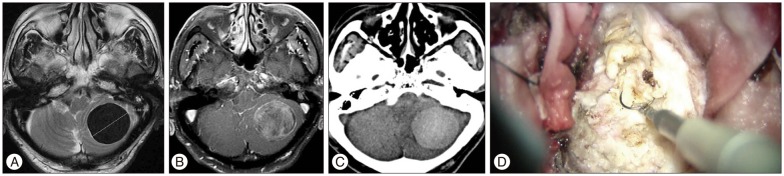
Fig. 1 Illustrative case. A and B : Preoperative MR images revealed 4 cm size round mass in cerebellum which was very low signal intensity on T2-weighted image without peritumoral edema and heterogeneously enhancing lesion after contrast administration. C : Preoperative CT scan showed high attenuated lesion with diffuse and scattered calcification. D : Intraoperative photograph shows entirely avascular nature with yellowish color and clear margin to brain parenchyme. Because the tumor was very tough and hard, the tumor was removed totally by using an electromagnetic field system.
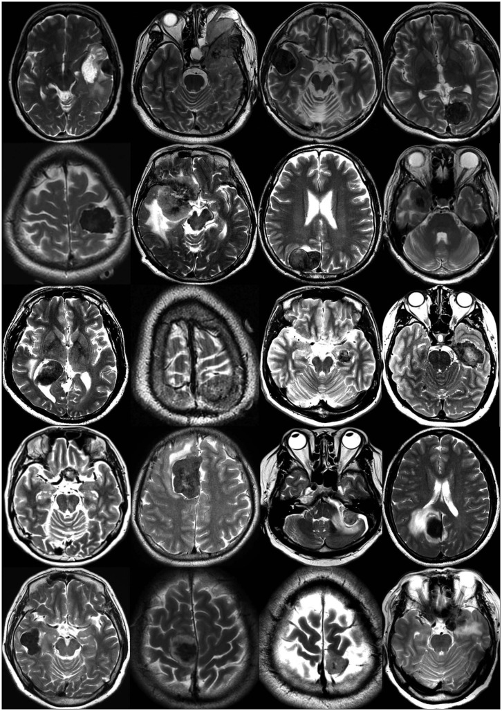
Fig. 2 T2-wheighted MR images of all patients with sclerosing meningioma except for the illustrative case. Among 21 patients, 15 patients (71%) have their distinctive very low signal intensity which seemed to be nearly dark color on T2-weighted images whereas isodense signal intensity on T2 images was found in only 6 patients. Furthermore, the majority of sclerosing meningioma revealed absent or minimal extent of peritumoral edema. Moderate to severe peritumoral edema found in only 4 patients.
Cited by 1 articles
-
Intracranial Metaplastic Meningioma : Clinical and Radiological Characteristics of 11 Cases
Taehoon Kim, Jin Wook Kim, So Young Ji, Ho Kang, Kyung-Min Kim, Yong Hwy Kim, Chul-Kee Park, Seung Hong Choi, Sung-Hye Park
J Korean Neurosurg Soc. 2020;63(5):657-663. doi: 10.3340/jkns.2020.0151.
Reference
-
1. Davidson GS, Hope JK. Meningeal tumors of childhood. Cancer. 1989; 63:1205–1210. PMID: 2645039.
Article2. Earnest F 4th, Kelly PJ, Scheithauer BW, Kall BA, Cascino TL, Ehman RL, et al. Cerebral astrocytomas : histopathologic correlation of MR and CT contrast enhancement with stereotactic biopsy. Radiology. 1988; 166:823–827. PMID: 2829270.
Article3. Elster AD, Challa VR, Gilbert TH, Richardson DN, Contento JC. Meningiomas : MR and histopathologic features. Radiology. 1989; 170(3 Pt 1):857–862. PMID: 2916043.4. Fukushima S, Narita Y, Yonezawa M, Ohno M, Arita H, Miyakita Y, et al. Short communication : sclerosing meningioma in the deep sylvian fissure. Brain Tumor Pathol. 2014; 31:289–292. PMID: 24141724.
Article5. Haberler C, Jarius C, Lang S, Rössler K, Gruber A, Hainfellner JA, et al. Fibrous meningeal tumours with extensive non-calcifying collagenous whorls and glial fibrillary acidic protein expression : the whorling-sclerosing variant of meningioma. Neuropathol Appl Neurobiol. 2002; 28:42–47. PMID: 11849562.
Article6. Hope JK, Armstrong DA, Babyn PS, Humphreys RR, Harwood-Nash DC, Chuang SH, et al. Primary meningeal tumors in children : correlation of clinical and CT findings with histologic type and prognosis. AJNR Am J Neuroradiol. 1992; 13:1353–1364. PMID: 1414828.7. Im SH, Chung CK, Cho BK, Kim MK, Chi JG. Sclerosing meningioma : clinicopathological study of four cases. J Neurooncol. 2004; 68:169–175. PMID: 15218954.
Article8. Kaplan RD, Coons S, Drayer BP, Bird CR, Johnson PC. MR characteristics of meningioma subtypes at 1.5 tesla. J Comput Assist Tomogr. 1992; 16:366–371. PMID: 1592917.
Article9. Kim BW, Kim MS, Kim SW, Chang CH, Kim OL. Peritumoral brain edema in meningiomas : correlation of radiologic and pathologic features. J Korean Neurosurg Soc. 2011; 49:26–30. PMID: 21494359.
Article10. Kim JW, Park CK, Park SH, Kim YH, Han JH, Kim CY, et al. Relationship between radiological characteristics and combined 1p and 19q deletion in World Health Organization grade III oligodendroglial tumours. J Neurol Neurosurg Psychiatry. 2011; 82:224–227. PMID: 20587495.
Article11. Kim NR, Im SH, Chung CK, Suh YL, Choe G, Chi JG. Sclerosing meningioma : immunohistochemical analysis of five cases. Neuropathol Appl Neurobiol. 2004; 30:126–135. PMID: 15043710.12. Kollová A, Liscák R, Novotný J Jr, Vladyka V, Simonová G, Janousková L. Gamma Knife surgery for benign meningioma. J Neurosurg. 2007; 107:325–336. PMID: 17695387.
Article13. Maiuri F, Iaconetta G, de Divitiis O, Cirillo S, Di Salle F, De Caro ML. Intracranial meningiomas : correlations between MR imaging and histology. Eur J Radiol. 1999; 31:69–75. PMID: 10477102.14. Megyesi JF, Kachur E, Lee DH, Zlatescu MC, Betensky RA, Forsyth PA, et al. Imaging correlates of molecular signatures in oligodendrogliomas. Clin Cancer Res. 2004; 10:4303–4306. PMID: 15240515.
Article15. Paek SH, Kim SH, Chang KH, Park CK, Kim JE, Kim DG, et al. Microcystic meningiomas : radiological characteristics of 16 cases. Acta Neurochir (Wien). 2005; 147:965–972. discussion 972. PMID: 16028111.
Article16. Pope LZ, Tatsui CE, Moro MS, Neto AC, Bleggi-Torres LF. Meningioma with extensive noncalcifying collagenous whorls and glial fibrillary acidic protein expression : new variant of meningioma diagnosed by smear preparation. Diagn Cytopathol. 2003; 28:274–277. PMID: 12722124.
Article17. Sheikh BY, Siqueira E, Dayel F. Meningioma in children : a report of nine cases and a review of the literature. Surg Neurol. 1996; 45:328–335. PMID: 8607080.
Article18. Stevens JM, Ruiz JS, Kendall BE. Observation on peritumoural oedema in meningioma. Part I : distribution, spread and resolution of vasogenic oedema seen on computed tomography. Neuroradiology. 1983; 25:71–80. PMID: 6877589.19. Walker DG, Kaye AH. Diagnosis and management of astrocytomas, oligodendrogliomas and mixed gliomas : a review. Australas Radiol. 2001; 45:472–482. PMID: 11903181.
Article20. Zlatescu MC, TehraniYazdi A, Sasaki H, Megyesi JF, Betensky RA, Louis DN, et al. Tumor location and growth pattern correlate with genetic signature in oligodendroglial neoplasms. Cancer Res. 2001; 61:6713–6715. PMID: 11559541.
- Full Text Links
-
- Actions
-
Cited
- CITED
-
- Close
- Share
-
- Similar articles
-
- Intracranial Metaplastic Meningioma : Clinical and Radiological Characteristics of 11 Cases
- A Case of Totally Calcified Meningioma
- The malignant meningioma with extracranial metastasis
- Meningioma of the Frontal and Ethmoidal Sinus: Case Report
- Ewing's Sarcoma Mimicking a Meningioma in Radiological Findings: A Case Report
