Obstet Gynecol Sci.
2018 May;61(3):367-373. 10.5468/ogs.2018.61.3.367.
Sonographic evaluation of bladder wall thickness in women with lower urinary tract dysfunction
- Affiliations
-
- 1Department of Obstetrics and Gynecology, University of Ulsan College of Medicine, Asan Medical Center, Seoul, Korea. hdchae@amc.seoul.kr
- 2Department of Obstetrics and Gynecology, Jeju National University College of Medicine, Jeju National University Hospital, Jeju, Korea.
- 3Department of Obstetrics and Gynecology, Eulji University, Nowon Eulji Medical Center, Seoul, Korea.
- KMID: 2416125
- DOI: http://doi.org/10.5468/ogs.2018.61.3.367
Abstract
OBJECTIVE
To investigate the correlation between bladder wall thickness (BWT) measured by ultrasonography and lower urinary tract dysfunction (LUTD) in patients with lower urinary tract symptoms (LUTS).
METHODS
Forty-eight women with LUTS who underwent urodynamic study and BWT by ultrasonography as outpatients were studied. We assessed LUTS during a medical examination by interview. The thinnest part of the bladder wall was measured by a transabdominal ultrasonography. We excluded patients who had visited another hospital previously because we did not know what treatment they had received, including medications, behavioral therapy, or other treatments. We constructed receiver operating characteristic (ROC) curves for diagnosis of LUTD and also determined reliable BWT criteria by calculating the area under the curve. Statistical analyses were performed using the Kolmogorov-Smirnov method and Student's t-test.
RESULTS
The mean age, body mass index, and duration of symptoms were 59.9±9.7 years, 26.06±3.4 kg/m², and 53.4±38.2 months, respectively. Urodynamic study parameters (Valsalva leak point pressure, maximal urethral closure pressure, functional length, and postvoid residual volume) were lower in patients with BWT < 3 mm; however, these differences were not significant. Patients with BWT ≥3 mm developed a hypoactive bladder (P=0.009) and intrinsic sphincter deficiency (ISD) (P=0.001) at a significantly higher rate. According to the ROC analysis, the best BWT cut-off value was 3 mm for overactive bladder diagnosis.
CONCLUSIONS
Women with LUTD showed higher BWT values (≥3 mm), especially patients with hypoactive bladder and ISD. Sonographic evaluation of BWT is an easy, fast, and noninvasive method for possible diagnostic tool for LUTD.
MeSH Terms
Figure
-
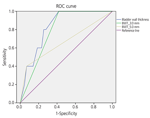
Fig. 1 Receiver operating characteristic (ROC) curve of the relationship between specificity and sensitivity for symptomatically diagnosed overactive bladder (OAB) patients according to bladder wall thickness (BWT).
Reference
-
1. Lee YS, Lee KS, Jung JH, Han DH, Oh SJ, Seo JT, et al. Prevalence of overactive bladder, urinary incontinence, and lower urinary tract symptoms: results of Korean EPIC study. World J Urol. 2011; 29:185–190.
Article2. Abrams P, Cardozo L, Fall M, Griffiths D, Rosier P, Ulmsten U, et al. The standardisation of terminology of lower urinary tract function: report from the Standardisation Sub-committee of the International Continence Society. Neurourol Urodyn. 2002; 21:167–178.
Article3. Ke QS, Kuo HC. The promise of bladder wall thickness as a useful biomarker for objective diagnosis of lower urinary tract dysfunction. Ci Ji Yi Xue Za Zhi. 2011; 23:1–8.
Article4. Ubee SS, Manikandan R, Singh G. Medical management of overactive bladder. Indian J Urol. 2010; 26:270–278.
Article5. Winters JC, Dmochowski RR, Goldman HB, Herndon CD, Kobashi KC, Kraus SR, et al. Urodynamic studies in adults: AUA/SUFU guideline. J Urol. 2012; 188:2464–2472.
Article6. Onur R, Ozden M, Orhan I, Kalkan A, Semercioz A. Incidence of bacteraemia after urodynamic study. J Hosp Infect. 2004; 57:241–244.
Article7. Güzel Ö, Aslan Y, Balcı M, Tuncel A, Keten T, Erkan A, et al. Can bladder wall thickness measurement be used for detecting bladder outlet obstruction? Urology. 2015; 86:439–444.
Article8. Ali MM, Ahmed AF, Khaled SM, Abozeid H, AbdelMagid ME. Accuracy of ultrasound-measured bladder wall thickness for the diagnosis of detrusor overactivity. Afr J Urol. 2015; 21:25–29.
Article9. Blatt AH, Titus J, Chan L. Ultrasound measurement of bladder wall thickness in the assessment of voiding dysfunction. J Urol. 2008; 179:2275–2278.
Article10. Üçer O, Gümüş B, Albaz AC, Pekindil G. Assessment of bladder wall thickness in women with overactive bladder. Turk J Urol. 2016; 42:97–100.11. Cruz F, Heesakkers J, Khullar V, Tubaro A. Bladder wall thickness in overactive bladder: does it have a role? Eur Urol Suppl. 2009; 8:769–771.
Article12. Lekskulchai O, Dietz HP. Detrusor wall thickness as a test for detrusor overactivity in women. Ultrasound Obstet Gynecol. 2008; 32:535–539.
Article13. Oelke M. International Consultation on Incontinence-Research Society (ICI-RS) report on non-invasive urodynamics: the need of standardization of ultrasound bladder and detrusor wall thickness measurements to quantify bladder wall hypertrophy. Neurourol Urodyn. 2010; 29:634–639.
Article14. Oelke M, Hofner K, Jonas U, Ubbink D, de la Rosette J, Wijkstra H. Ultrasound measurement of detrusor wall thickness in healthy adults. Neurourol Urodyn. 2006; 25:308–317.
Article15. Dietz HP. Ultrasound imaging of the pelvic floor. Part 1: two-dimensional aspects. Ultrasound Obstet Gynecol. 2004; 23:80–92.16. Otsuki EN, Araujo Júnior E, Oliveira E, Sartori MG, Girão MJ, Jármy-Di Bella ZI. Ultrasound thickness of bladder wall in continent and incontinent women and its correlation with cystometry. ScientificWorldJournal. 2014; 2014:684671.
Article17. Robinson D, Anders K, Cardozo L, Bidmead J, Toozs-Hobson P, Khullar V. Can ultrasound replace ambulatory urodynamics when investigating women with irritative urinary symptoms? BJOG. 2002; 109:145–148.
Article18. Kojima M, Inui E, Ochiai A, Naya Y, Ukimura O, Watanabe H. Noninvasive quantitative estimation of infravesical obstruction using ultrasonic measurement of bladder weight. J Urol. 1997; 157:476–479.
Article19. Kuhn A, Genoud S, Robinson D, Herrmann G, Günthert A, Brandner S, et al. Sonographic transvaginal bladder wall thickness: does the measurement discriminate between urodynamic diagnoses? Neurourol Urodyn. 2011; 30:325–328.
Article20. Serati M, Salvatore S, Cattoni E, Soligo M, Cromi A, Ghezzi F. Ultrasound measurement of bladder wall thickness in different forms of detrusor overactivity. Int Urogynecol J Pelvic Floor Dysfunct. 2010; 21:1405–1411.
Article21. Pannek J, Bartel P, Gocking K, Frotzler A. Clinical usefulness of ultrasound assessment of detrusor wall thickness in patients with neurogenic lower urinary tract dysfunction due to spinal cord injury: urodynamics made easy? World J Urol. 2013; 31:659–664.
Article22. Miyazato M, Yoshimura N, Chancellor MB. The other bladder syndrome: underactive bladder. Rev Urol. 2013; 15:11–22.23. Diokno AC, Rashid TM. The artificial urinary sphincter. In : Stanton SL, Zimmern P, editors. Female pelvic reconstructive surgery. 3rd ed. London: Springer;2005. p. 141–147.
- Full Text Links
-
- Actions
-
Cited
- CITED
-
- Close
- Share
-
- Similar articles
-
- Pathophysiology of lower urinary tract dysfunction in the older patient
- Increased Bladder Wall Thickness in Diabetic and Nondiabetic Women With Overactive Bladder
- Changes in Bladder Wall Thickness and Detrusor Wall Thickness After Surgical Treatment of Benign Prostatic Enlargement in Patients With Lower Urinary Tract Symptoms: A Preliminary Report
- Leiomyosarcoma of the Urinary Bladder
- Overview of the Epidemiology of Lower Urinary Tract Dysfunction in South Korea
