Stress Fracture of the Capitate
- Affiliations
-
- 1Department of Radiology, Korea University Anam Hospital, Seoul, Korea.
- 2Department of Radiology, Korea University College of Medicine, Seoul, Korea. keytech2@naver.com
- KMID: 2415894
- DOI: http://doi.org/10.13104/imri.2018.22.2.135
Abstract
- Most capitate fractures occur in association with additional carpal injuries, particularly scaphoid fractures. Isolated fractures of the capitate account for only 0.3% of carpal injuries, and stress fractures are one form of this fracture. We report the case of a 20-year-old male who had a stress fracture of the capitate after serving as an honor guard in the military. Conventional radiographs and computed tomography of the right wrist revealed a minimally displaced fracture line located at the midcarpal aspect of the right capitate. A magnetic resonance imaging scan demonstrates a subarticular capitate fracture with diffuse bone marrow edema, small osteophytes, and irregularity of the midcarpal articular cartilage. We also review the carpal kinematics which possibly caused the stress fracture. Although stress fractures of the capitate are rare, they should also be accounted for with patients who perform repetitive motions of the wrist to a considerable extent.
Keyword
MeSH Terms
Figure
-
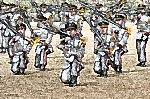
Fig. 1 An illustration of the honor guard during the ceremony.
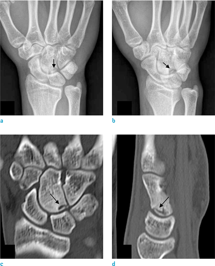
Fig. 2 Radiographs and CT of the right wrist of a 20-year-old honor guard suffering stress fracture of the capitate. (a, b) Anteroposterior and oblique radiographs demonstrate the fracture line located at the midcarpal aspect of the right capitate (arrows). (c, d) Coronal and sagittal multiplanar reconstruction bone window CT showed a minimally displaced curved fracture with surrounding sclerotic change (arrows).
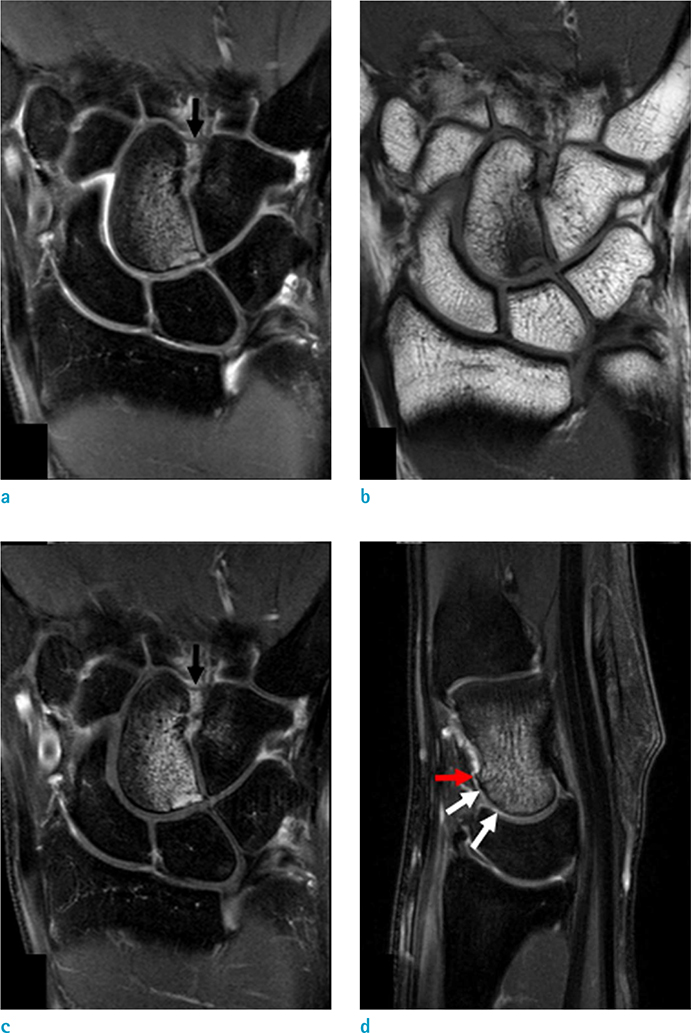
Fig. 3 (a-c) Coronal proton-density-weighted fat-saturated, T1-weighted, and contrast agent-enhanced T1-weighted fat-saturated MR images of the right wrist show subarticular capitate fracture with diffuse bone marrow edema. An increased signal intensity with thickened volar capito-hamate ligament is also witnessed, which indicates a suspicious ligament injury (black arrows). (d) Sagittal T2-weighted fat-saturated MR image demonstrates small osteophytes in the capitate (red arrow) with irregularity of the midcarpal articular cartilage (white arrows), suggesting osteoarthritis.
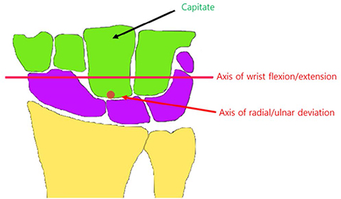
Fig. 4 Kinematics of the wrist during flexion-extension and radioulnar deviation, with two axes of motion located within the head of the capitate.
Reference
-
1. Panchal-Kildare S, Malone K. Skeletal anatomy of the hand. Hand Clin. 2013; 29:459–471.
Article2. Kaewlai R, Avery LL, Asrani AV, Abujudeh HH, Sacknoff R, Novelline RA. Multidetector CT of carpal injuries: anatomy, fractures, and fracture-dislocations. Radiographics. 2008; 28:1771–1784. abstract vii.
Article3. Vigler M, Aviles A, Lee SK. Carpal fractures excluding the scaphoid. Hand Clin. 2006; 22:501–516.
Article4. Oestreich AE, Bhojwani N. Stress fractures of ankle and wrist in childhood: nature and frequency. Pediatr Radiol. 2010; 40:1387–1389.
Article5. Jaimes C, Jimenez M, Shabshin N, Laor T, Jaramillo D. Taking the stress out of evaluating stress injuries in children. Radiographics. 2012; 32:537–555.
Article6. Vizkelety T, Wouters HW. Stress fracture of the capitate. Arch Chir Neerl. 1972; 24:47–57.7. Allen H, Gibbon WW, Evans RJ. Stress fracture of the capitate. J Accid Emerg Med. 1994; 11:59–60.
Article8. Moojen TM, Snel JG, Ritt MJ, Venema HW, Kauer JM, Bos KE. In vivo analysis of carpal kinematics and comparative review of the literature. J Hand Surg Am. 2003; 28:81–87.
Article9. Moritomo H, Murase T, Goto A, Oka K, Sugamoto K, Yoshikawa H. Capitate-based kinematics of the midcarpal joint during wrist radioulnar deviation: an in vivo three-dimensional motion analysis. J Hand Surg Am. 2004; 29:668–675.
Article10. Berger FH, de Jonge MC, Maas M. Stress fractures in the lower extremity. The importance of increasing awareness amongst radiologists. Eur J Radiol. 2007; 62:16–12.
- Full Text Links
-
- Actions
-
Cited
- CITED
-
- Close
- Share
-
- Similar articles
-
- Fracture of the Capitate with Velar Perilunate Dislocation: One case report
- Avascular Necrosis of the Capitate: MRI and Surgical Decision-Making of Two Case Reports
- Idiopathic Avascular Necrosis of the Capitate: A Case Report
- Solitary Bone Cyst of the Capitate: A Case Report
- Subcapital Stress Fracture of the Femur after Internal Fixation of Intertrochanteric Fracture: A case report
