Ann Dermatol.
2018 Apr;30(2):247-249. 10.5021/ad.2018.30.2.247.
Fibrous Plaque of the Eyelid in a Patient with Tuberous Sclerosis Responding to Everolimus
- Affiliations
-
- 1Department of Dermatology, Pusan National University School of Medicine, Yangsan, Korea. hcko@pusan.ac.kr
- 2Department of Dermatology, Pusan National University Yangsan Hospital, Yangsan, Korea.
- 3Department of Pediatrics, Pusan National University School of Medicine, Yangsan, Korea.
- KMID: 2414696
- DOI: http://doi.org/10.5021/ad.2018.30.2.247
Abstract
- No abstract available.
Figure
-
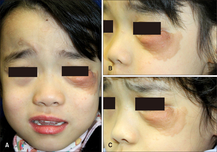
Fig. 1 (A, B) Erythematous indurated plaque of the left lower eyelid, skin-colored to brownish plaques on the forehead. (C) Decrease in thickness and erythema of the lesion after oral everolimus treatment for 14 months.
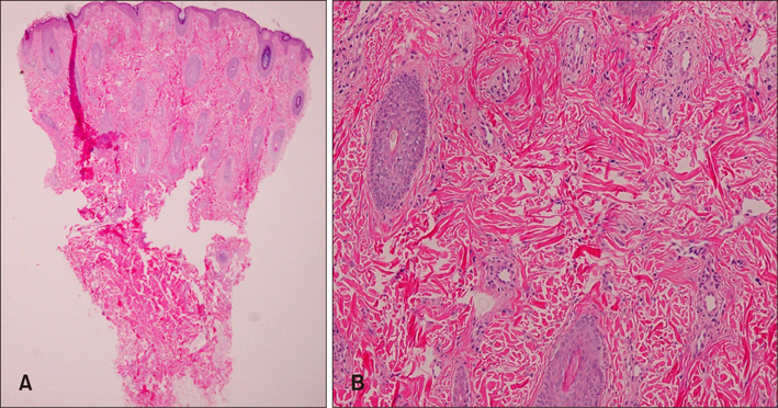
Fig. 2 Histological examination of the eyelid specimen shows fibrovascular proliferation and hyperplasia of hair follicles (H&E; A: ×20, B: ×100).
Reference
-
1. Jacks SK, Witman PM. Tuberous sclerosis complex: an update for dermatologists. Pediatr Dermatol. 2015; 32:563–570.
Article2. Torrelo A, Hadj-Rabia S, Colmenero I, Piston R, Sybert VP, Hilari-Carbonell H, et al. Folliculocystic and collagen hamartoma of tuberous sclerosis complex. J Am Acad Dermatol. 2012; 66:617–621.
Article3. Nathan N, Wang JA, Li S, Cowen EW, Haughey M, Moss J, et al. Improvement of tuberous sclerosis complex (TSC) skin tumors during long-term treatment with oral sirolimus. J Am Acad Dermatol. 2015; 73:802–808.
Article4. Karar J, Maity A. PI3K/AKT/mTOR pathway in angiogenesis. Front Mol Neurosci. 2011; 4:51.
Article5. Zolli C, Rodrigues MM, Shannon GM. Unusual eyelid involvement in tuberous sclerosis. J Pediatr Ophthalmol. 1976; 13:156–158.
Article
- Full Text Links
-
- Actions
-
Cited
- CITED
-
- Close
- Share
-
- Similar articles
-
- A Case of Forehead Plaque as an Initial Sign of Tuberous Sclerosis
- Erratum: Fibrous Plaque of the Eyelid in a Patient with Tuberous Sclerosis Responding to Everolimus
- Everolimus Treatment and Follow-up in an Infant with Neurological Complication Associated with Tuberous Sclerosis Complex
- Tuberous Sclerosis with Aortic Aneurysm and Rib Changes
- Experience of a Single Center in Treating Multiple Manifestations of Tuberous Sclerosis Complex with Everolimus
