Computer-Aided Diagnosis of Thyroid Nodules via Ultrasonography: Initial Clinical Experience
- Affiliations
-
- 1Department of Radiology, Ajou University School of Medicine, Suwon 16499, Korea. radhej@naver.com
- 2Department of Biostatistics, Ajou University School of Medicine, Suwon 16499, Korea.
- KMID: 2413695
- DOI: http://doi.org/10.3348/kjr.2018.19.4.665
Abstract
OBJECTIVE
To prospectively evaluate the diagnostic performance of computer-aided diagnosis (CAD) for detection of thyroid cancers via ultrasonography (US).
MATERIALS AND METHODS
This study included 50 consecutive patients with 117 thyroid nodules on US during the period between June 2016 and July 2016. A radiologist performed US examinations using real-time CAD integrated into a US scanner. We compared the diagnostic performance of radiologist, the CAD system, and the CAD-assisted radiologist for the detection of thyroid cancers.
RESULTS
The sensitivity, specificity, positive predictive value (PPV), negative predictive value (NPV), and accuracy of the CAD system were 80.0, 88.1, 83.3, 85.5, and 84.6%, respectively, and were not significantly different from those of the radiologist (p > 0.05). The CAD-assisted radiologist showed improved diagnostic sensitivity compared with the radiologist alone (92.0% vs. 84.0%, p = 0.037), while the specificity and PPV were reduced (85.1% vs. 95.5%, p = 0.005 and 82.1% vs. 93.3%, p = 0.008). The radiologist assisted by the CAD system exhibited better diagnostic sensitivity and NPV than the CAD system alone (92.0% vs. 80.0%, p = 0.009 and 93.4% vs. 88.9%, p = 0.013), while the specificities and PPVs were not significantly different (88.1% vs. 85.1%, p = 0.151 and 83.3% vs. 82.1%, p = 0.613, respectively).
CONCLUSION
The CAD system may be an adjunct to radiological intervention in the diagnosis of thyroid cancer.
Keyword
MeSH Terms
Figure
-
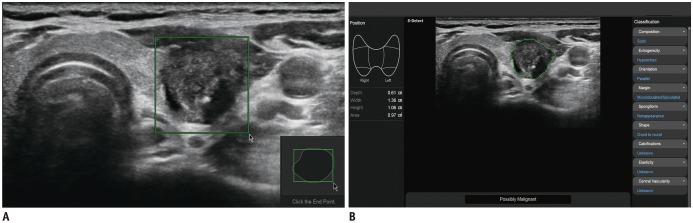
Fig. 1 US image of thyroid nodule acquired via CAD system.A. Solid hypoechoic nodule with suspicious US features is evident in left thyroid gland. Region of interest is manually drawn around lesion. B. CAD software automatically calculates mass contours and presents US features on right of screen, and possible diagnosis as malignant nodule at bottom. CAD = computer-aided diagnosis, US = ultrasonography
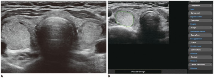
Fig. 2 53-year-old woman with bilateral thyroid nodules.A. US images show solid isoechoic nodule without suspicious US features in right thyroid gland. Radiological diagnosis suggested benign nodule. B. CAD system presented possible diagnosis of benign nodule. Histology confirmed adenomatous hyperplasia.
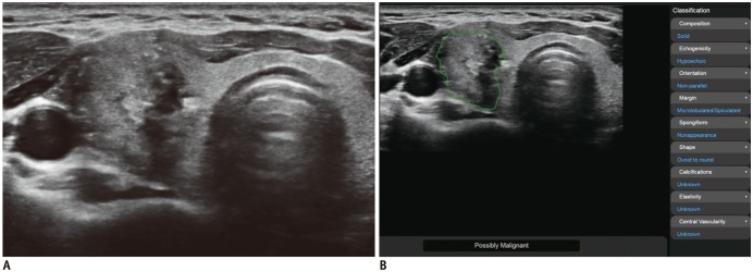
Fig. 3 47-year-old woman with right thyroid nodule.A. US images show solid hypoechoic nodule with suspicious US features in right thyroid gland. Radiological diagnosis suggested malignant nodule. B. CAD system presented possible diagnosis as malignant nodule. Histology confirmed diagnosis of papillary thyroid carcinoma.
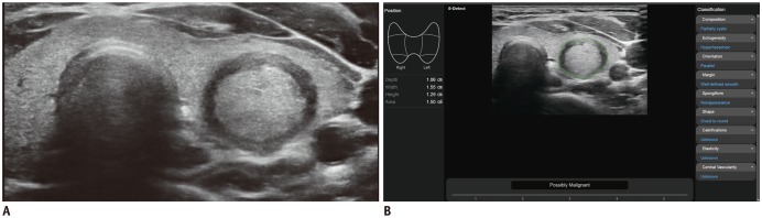
Fig. 4 36-year-old woman with left thyroid nodule.A. US images show solid isoechoic nodule with thick peripheral halo in left thyroid gland. Radiologist diagnosed it as benign nodule. B. CAD system suggested possible diagnosis of malignant nodule following US misdiagnosis. Histology confirmed diagnosis of adenomatous hyperplasia.
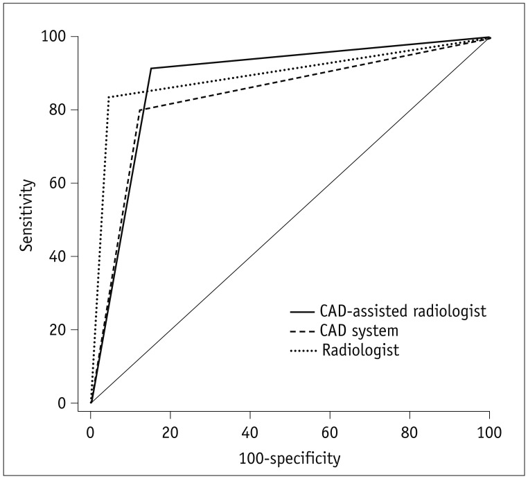
Fig. 5 Comparison of receiver operating characteristic curves for CAD, radiologist, and CAD-assisted radiologist in thyroid cancer diagnosis.
Cited by 2 articles
-
Computer-Aided Diagnosis System for the Evaluation of Thyroid Nodules on Ultrasonography: Prospective Non-Inferiority Study according to the Experience Level of Radiologists
Sae Rom Chung, Jung Hwan Baek, Min Kyoung Lee, Yura Ahn, Young Jun Choi, Tae-Yon Sung, Dong Eun Song, Tae Yong Kim, Jeong Hyun Lee
Korean J Radiol. 2020;21(3):369-376. doi: 10.3348/kjr.2019.0581.Clinical validation of a deep-learning-based bone age software in healthy Korean children
Hyo-Kyoung Nam, Winnah Wu-In Lea, Zepa Yang, Eunjin Noh, Young-Jun Rhie, Kee-Hyoung Lee, Suk-Joo Hong
Ann Pediatr Endocrinol Metab. 2024;29(2):102-108. doi: 10.6065/apem.2346050.025.
Reference
-
1. Brander A, Viikinkoski P, Nickels J, Kivisaari L. Thyroid gland: US screening in a random adult population. Radiology. 1991; 181:683–687. PMID: 1947082.
Article2. Haugen BR. 2015 American Thyroid Association management guidelines for adult patients with thyroid nodules and differentiated thyroid cancer: what is new and what has changed? Cancer. 2017; 123:372–381. PMID: 27741354.
Article3. Camacho PM, Petak SM, Binkley N, Clarke BL, Harris ST, Hurley DL, et al. American Association of Clinical Endocrinologists and American College of Endocrinology clinical practice guidelines for the diagnosis and treatment of postmenopausal osteoporosis - 2016. Endocr Pract. 2016; 22(Suppl 4):1–42.
Article4. Shin JH, Baek JH, Chung J, Ha EJ, Kim JH, Lee YH, et al. Korean Society of Thyroid Radiology (KSThR) and Korean Society of Radiology. Ultrasonography diagnosis and imaging-based management of thyroid nodules: revised Korean Society of Thyroid Radiology consensus statement and recommendations. Korean J Radiol. 2016; 17:370–395. PMID: 27134526.
Article5. Ko SY, Kim EK, Sung JM, Moon HJ, Kwak JY. Diagnostic performance of ultrasound and ultrasound elastography with respect to physician experience. Ultrasound Med Biol. 2014; 40:854–863. PMID: 24315394.
Article6. Kim HG, Kwak JY, Kim EK, Choi SH, Moon HJ. Man to man training: can it help improve the diagnostic performances and interobserver variabilities of thyroid ultrasonography in residents? Eur J Radiol. 2012; 81:e352–e356. PMID: 22137098.
Article7. Choi SH, Kim EK, Kwak JY, Kim MJ, Son EJ. Interobserver and intraobserver variations in ultrasound assessment of thyroid nodules. Thyroid. 2010; 20:167–172. PMID: 19725777.
Article8. Park CS, Kim SH, Jung SL, Kang BJ, Kim JY, Choi JJ, et al. Observer variability in the sonographic evaluation of thyroid nodules. J Clin Ultrasound. 2010; 38:287–293. PMID: 20544863.
Article9. Park SH, Kim SJ, Kim EK, Kim MJ, Son EJ, Kwak JY. Interobserver agreement in assessing the sonographic and elastographic features of malignant thyroid nodules. AJR Am J Roentgenol. 2009; 193:W416–W423. PMID: 19843721.
Article10. Park SJ, Park SH, Choi YJ, Kim DW, Son EJ, Lee HS, et al. Interobserver variability and diagnostic performance in US assessment of thyroid nodule according to size. Ultraschall Med. 2012; 33:E186–E190. PMID: 23108925.
Article11. Lim KJ, Choi CS, Yoon DY, Chang SK, Kim KK, Han H, et al. Computer-aided diagnosis for the differentiation of malignant from benign thyroid nodules on ultrasonography. Acad Radiol. 2008; 15:853–858. PMID: 18572120.
Article12. Chang Y, Paul AK, Kim N, Baek JH, Choi YJ, Ha EJ, et al. Computer-aided diagnosis for classifying benign versus malignant thyroid nodules based on ultrasound images: a comparison with radiologist-based assessments. Med Phys. 2016; 43:554. PMID: 26745948.
Article13. Li LN, Ouyang JH, Chen HL, Liu DY. A computer aided diagnosis system for thyroid disease using extreme learning machine. J Med Syst. 2012; 36:3327–3337. PMID: 22327384.
Article14. Acharya UR, Faust O, Sree SV, Molinari F, Garberoglio R, Suri JS. Cost-effective and non-invasive automated benign and malignant thyroid lesion classification in 3D contrast-enhanced ultrasound using combination of wavelets and textures: a class of ThyroScan™ algorithms. Technol Cancer Res Treat. 2011; 10:371–380. PMID: 21728394.15. Acharya UR, Faust O, Sree SV, Molinari F, Suri JS. ThyroScreen system: high resolution ultrasound thyroid image characterization into benign and malignant classes using novel combination of texture and discrete wavelet transform. Comput Methods Programs Biomed. 2012; 107:233–241. PMID: 22054816.
Article16. Acharya UR, Sree SV, Krishnan MM, Molinari F, Zieleźnik W, Bardales RH, et al. Computer-aided diagnostic system for detection of Hashimoto thyroiditis on ultrasound images from a Polish population. J Ultrasound Med. 2014; 33:245–253. PMID: 24449727.
Article17. Acharya UR, Vinitha Sree S, Krishnan MM, Molinari F, Garberoglio R, Suri JS. Non-invasive automated 3D thyroid lesion classification in ultrasound: a class of ThyroScan™ systems. Ultrasonics. 2012; 52:508–520. PMID: 22154208.
Article18. Choi YJ, Baek JH, Park HS, Shim WH, Kim TY, Shong YK, et al. A computer-aided diagnosis system using artificial intelligence for the diagnosis and characterization of thyroid nodules on ultrasound: initial clinical assessment. Thyroid. 2017; 27:546–552. PMID: 28071987.
Article
- Full Text Links
-
- Actions
-
Cited
- CITED
-
- Close
- Share
-
- Similar articles
-
- Sonographic Evaluation of Thyroid Nodules
- Computer-Aided Diagnosis System for the Evaluation of Thyroid Nodules on Ultrasonography: Prospective Non-Inferiority Study according to the Experience Level of Radiologists
- Applications of machine learning and deep learning to thyroid imaging: where do we stand?
- A Computer-Aided Diagnosis for Evaluating Lung Nodules on Chest CT: the Current Status and Perspective
- Natural Course of Cytologically Diagnosed Benign Thyroid Nodules
