Diagnostic imaging features of normal anal sacs in dogs and cats
- Affiliations
-
- 1Veterinary Clinical Sciences, College of Veterinary Medicine, Chonbuk National University, Iksan 54596, Korea. kclee@jbnu.ac.kr
- KMID: 2413132
- DOI: http://doi.org/10.4142/jvs.2016.17.3.331
Abstract
- This study was conducted to provide normal reference features for canine and feline anal sacs using ultrasound, low-field magnetic resonance imaging (MRI) and radiograph contrast as diagnostic imaging tools. A total of ten clinically normal beagle dogs and eight clinically normally cats were included. General radiography with contrast, ultrasonography and low-field MRI scans were performed. The visualization of anal sacs, which are located at distinct sites in dogs and cats, is possible with a contrast study on radiography. Most surfaces of the anal sacs tissue, occasionally appearing as a hyperechoic thin line, were surrounded by the hypoechoic external sphincter muscle on ultrasonography. The normal anal sac contents of dogs and cats had variable echogenicity. Signals of anal sac contents on low-field MRI varied in cats and dogs, and contrast medium using T1-weighted images enhanced the anal sac walls more obviously than that on ultrasonography. In conclusion, this study provides the normal features of anal sacs from dogs and cats on diagnostic imaging. Further studies including anal sac evaluation are expected to investigate disease conditions.
MeSH Terms
Figure
-
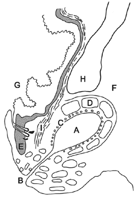
Fig. 1 Diagram of a dorsal section of the anal sac of a dog. (A) Anal sac cavity. (B) Anal sac duct opening. (C) Apocrine glands. (D) External sphincter muscle. (E) Internal sphincter muscle. (F) Fat of rectoischial fossa. (G) Anal canal. (H) Levator ani muscle. (I) Longitudinal muscle layer of rectum [15].
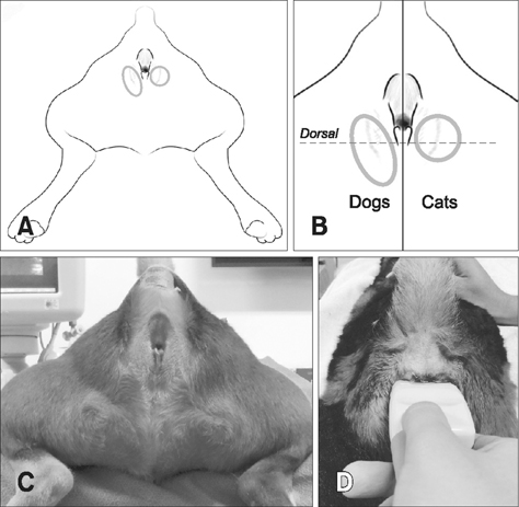
Fig. 2 Positioning for anal sac scan (A and C). Approaching for ultrasonographic examination of the anal sac (B and D). The dog (C) and cat (D) were sedated, but sedation was not essential.
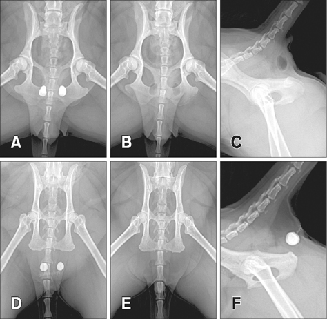
Fig. 3 Ventrodorsal (A, B, D and E) and lateral (C and F) view on radiograph of canine (A–C) and feline (C, D and F) pelvic region. The oval-shaped anal sacs were superimposed over the ischial table region on positive (A) and negative (B and C) contrast study images in dogs. However, the round-shaped anal sacs were observed in the soft tissue of the caudal region of the pelvis on positive (D and F) and negative (E) contrast study images in cats. The anal sac duct was visible as a radiopaque line (F).
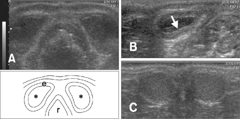
Fig. 4 Dorsal sonographic and diagram (A) of canine anal sac. Dorsal schematic sonograph image (B) of canine anal sac. Anal sac tissue (glands) appears as a hyperechoic thin line (arrow). Dorsal sonographic images of feline anal sac (C). The appearance of the anal sac was round, and their contents appear hypoechoic to similar to external sphincter muscle. Asterisks, anal sac contents; e, external sphincter muscle; r, rectum.
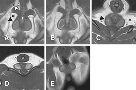
Fig. 5 Dorsal (A, B and E) and transverse (C and D) low-field MRI T1-weighted images of canine (A–C) and feline (D and E) anal sac. Increased signal intensity of anal sac tissue was identified on T1-weighted enhanced images (B). Asterisks, fat of ischiorectal fossa; arrow, levator ani muscle; arrow heads, external sphincter muscle; dagger, anal sac contents; r, rectum.
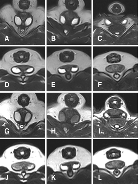
Fig. 6 Signals on T2-weighted (A–F) and FLAIR (G–L) images of canine (A–C and G–I) and feline (D–F and J–L) anal sac contents.
Reference
-
1. Al-bagdadi F. The integument. The digestive apparatus and abdomen. In : Evans HE, de Lahunta A, editors. Miller's Anatomy of the Dog. 4th ed. St. Louis: Saunders;2013. p. 75. p. 325.2. Aronson L. Rectum and anus. In : Slatter DH, editor. Textbook of Small Animal Surgery. 3rd ed. Philadelphia: Saunders;2003. p. 682–708.3. Bennett PF, DeNicola DB, Bonney P, Glickman NW, Knapp DW. Canine anal sac adenocarcinomas: clinical presentation and response to therapy. J Vet Intern Med. 2002; 16:100–104.
Article4. Brearley MJ. Epithelial and other solitary skin tumours. In : Dobson JM, Lascelles BDX, editors. BSAVA Manual of Canine and Feline Oncology. 2nd ed. Quedgeley: British Small Animal Veterinary Association;2003. p. 152–160.5. Craven M. Rectoanal disease. In : Ettinger SJ, Feldman EC, editors. Textbook of Veterinary Internal Medicine. 7th ed. St. Louis: Elsevier Saunders;2010. p. 1604–1606.6. Dennis R, Penderis J. Radiology corner—anal sac gas appearing as an osteolytic pelvic lesion. Vet Radiol Ultrasound. 2002; 43:552–553.
Article7. van Duijkeren E. Disease conditions of canine anal sacs. J Small Anim Pract. 1995; 36:12–16.
Article8. Elliott JW, Blackwood L. Treatment and outcome of four cats with apocrine gland carcinoma of the anal sac and review of the literature. J Feline Med Surg. 2011; 13:712–717.
Article9. Frankel JL, Scott DW, Erb HN. Gross and cytological characteristics of normal feline anal-sac secretions. J Feline Med Surg. 2008; 10:319–323.
Article10. Greer MB, Calhoun ML. Anal sacs of the cat (Felis domesticus). Am J Vet Res. 1966; 27:773–781.11. Halnan CR. The diagnosis of anal sacculitis in the dog. J Small Anim Pract. 1976; 17:527–535.
Article12. Harvey CE. Incidence distribution of anal sac disease in the dog. J Am Anim Hosp Assoc. 1974; 10:573–577.13. James DJ, Griffin CE, Polissar NL, Neradilek MB. Comparison of anal sac cytological findings and behaviour in clinically normal dogs and those affected with anal sac disease. Vet Dermatol. 2011; 22:80–87.
Article14. Lake AM, Scott DW, Miller WH Jr, Erb HN. Gross and cytological characteristics of normal canine anal-sac secretions. J Vet Med A Physiol Pathol Clin Med. 2004; 51:249–253.
Article15. McColl I. The comparative anatomy and pathology of anal glands. Arris and Gale lecture delivered at the Royal College of Surgeons of England on 25th February 1965. Ann R Coll Surg Engl. 1967; 40:36–67.16. Pappalardo E, Martino PA, Noli C. Macroscopic, cytological and bacteriological evaluation of anal sac content in normal dogs and in dogs with selected dermatological diseases. Vet Dermatol. 2002; 13:315–322.
Article
- Full Text Links
-
- Actions
-
Cited
- CITED
-
- Close
- Share
-
- Similar articles
-
- Isolation Rates and Carrier State of Dermatophytes, Nondermatophyte Molds, Malassezia Species, and Candida Species in Indoor Dogs and Cats in Daegu
- Guidelines for vaccination of dogs and cats in Korea
- Rabies neutralizing antibody titers in Korean dogs and cats intended for overseas travel
- Computed tomographic evaluation Medical Imaging of portal vein indices in cats with the extrahepatic portosystemic shunts
- Demographics of dogs and cats with oral tumors presenting to teaching hospitals: 1996–2017
