Merkel Cell Carcinoma Metastatic to Pleural Fluid: A Case Report
- Affiliations
-
- 1Pathology Center, Seegene Medical Foundation, Seoul, Korea.
- 2Department of Pathology, Severance Hospital, Yonsei University College of Medicine, Seoul, Korea. paxco@yuhs.ac
- KMID: 2412936
- DOI: http://doi.org/10.4132/jptm.2017.11.10
Abstract
- Merkel cell carcinoma (MCC) is a rare aggressive neuroendocrine carcinoma of the skin that shows locoregional or distant metastasis. Metastasis of MCC to body cavity effusion is extremely rare; only three cases have been reported so far. Metastatic MCC in effusion cytology shows small blue round cells with fine stippled chromatin like other small blue round cell tumors such as small cell lung carcinoma or lymphoma. The diagnosis of metastatic MCC can grant patients good chances at recently advanced therapeutic options. Here, we present a case of metastatic MCC to pleural effusion with characteristic single file-like pattern.
MeSH Terms
Figure
-

Fig. 1. Chest computed tomography (CT) with cell block findings. The chest CT shows marked pleural effusion (arrows).
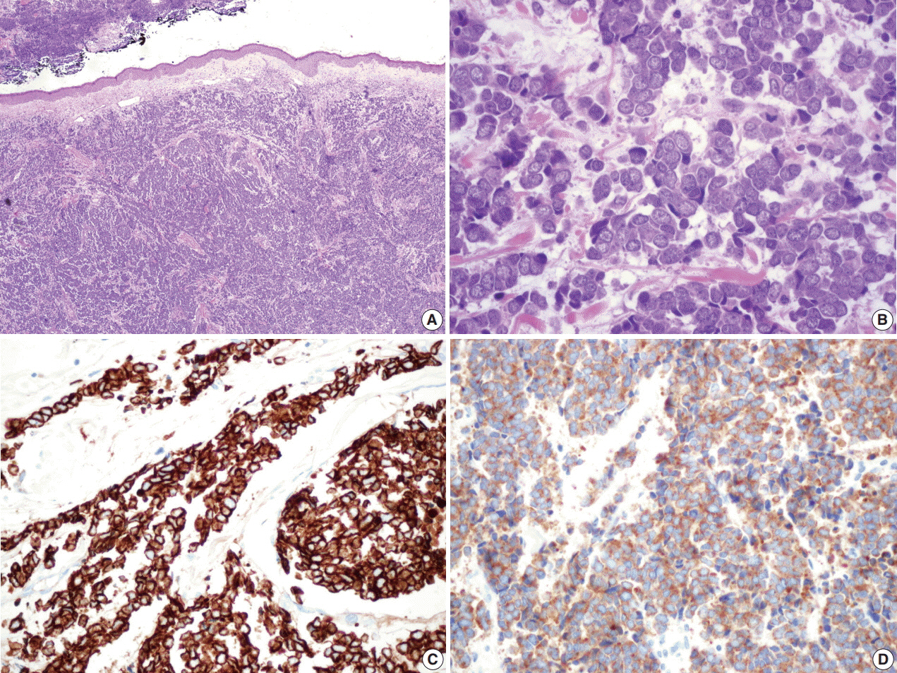
Fig. 2. Microscopic findings of the surgical specimen. Blue small round cell tumor with infiltrative growth (A) arranged in sheets, clusters, rows or balls, showing hyperchromatic nuclei, fine stippled or smudged chromatin and occasional mitoses (B). Immunohistochemical staining of cytokeratin 20 (C) and synaptophysin (D) show positive reaction in Merkel cell carcinoma cells.

Fig. 3. (A) Liquid based cytology (BD Surepath) of the pleural effusion (Papanicolaou stain). Malignant small blue round cells are scattered, clustered or arranged with hyperchromatic nuclei, fine granular salt and pepper chromatin, scant cytoplasm, and occasional mitotic figures. Several single file patterns with nuclear molding are characteristic (arrows). (B) The cell block findings are similar to those in liquid based cytology’s. Immunohistochemical staining of cytokeratin 20 (C) and synaptophysin (D) in the cell block of pleural effusion.
Reference
-
1. Hodgson NC. Merkel cell carcinoma: changing incidence trends. J Surg Oncol. 2005; 89:1–4.
Article2. Feng H, Shuda M, Chang Y, Moore PS. Clonal integration of a polyomavirus in human Merkel cell carcinoma. Science. 2008; 319:1096–100.
Article3. Pulitzer MP, Amin BD, Busam KJ. Merkel cell carcinoma: review. Adv Anat Pathol. 2009; 16:135–44.4. Toker C. Trabecular carcinoma of the skin. Arch Dermatol. 1972; 105:107–10.
Article5. Pulitzer M. Merkel cell carcinoma. Surg Pathol Clin. 2017; 10:399–408.
Article6. Watson CW, Friedman KJ. Cytology of metastatic neuroendocrine (Merkel-cell) carcinoma in pleural fluid: a case report. Acta Cytol. 1985; 29:397–402.7. Payne MM, Rader AE, McCarthy DM, Rodgers WH. Merkel cell carcinoma in a malignant pleural effusion: case report. Cytojournal. 2004; 1:5.8. Policarpio-Nicolas ML, Avery DL, Hartley T. Merkel cell carcinoma presenting as malignant ascites: a case report and review of literature. Cytojournal. 2015; 12:19.
Article9. Fitzgerald TL, Dennis S, Kachare SD, Vohra NA, Wong JH, Zervos EE. Dramatic increase in the incidence and mortality from Merkel cell carcinoma in the United States. Am Surg. 2015; 81:802–6.
Article10. Robertson JP, Liang ES, Martin RC. Epidemiology of Merkel cell carcinoma in New Zealand: a population-based study. Br J Dermatol. 2015; 173:835–7.
Article11. Eng TY, Boersma MG, Fuller CD, et al. A comprehensive review of the treatment of Merkel cell carcinoma. Am J Clin Oncol. 2007; 30:624–36.
Article12. Allen PJ, Bowne WB, Jaques DP, Brennan MF, Busam K, Coit DG. Merkel cell carcinoma: prognosis and treatment of patients from a single institution. J Clin Oncol. 2005; 23:2300–9.
Article13. Barksdale SK. Advances in Merkel cell carcinoma from a pathologist’s perspective. Pathology. 2017; 49:568–74.
Article
