Comparison of Anterior, Posterior, and Total Corneal Astigmatism Measured Using a Single Scheimpflug Camera in Healthy and Keratoconus Eyes
- Affiliations
-
- 1Department of Ophthalmology, Korea University College of Medicine, Seoul, Korea. hippotate@hanmail.net
- KMID: 2412794
- DOI: http://doi.org/10.3341/kjo.2017.0075
Abstract
- PURPOSE
To compare the effect of posterior corneal astigmatism on the estimation of total corneal astigmatism using anterior corneal measurements (simulated keratometry [K]) between eyes with keratoconus and healthy eyes.
METHODS
Thirty-three eyes of 33 patients with keratoconus of grade I or II and 33 eyes of 33 age- and sex-matched healthy control subjects were enrolled. Anterior, posterior, and total corneal cylinder powers and flat meridians measured by a single Scheimpflug camera were analyzed. The difference in corneal astigmatism between the simulated K and total cornea was evaluated.
RESULTS
The mean anterior, posterior, and total corneal cylinder powers of the keratoconus group (4.37 ± 1.73, 0.95 ± 0.39, and 4.36 ± 1.74 cylinder diopters [CD], respectively) were significantly greater than those of the control group (1.10 ± 0.68, 0.39 ± 0.18, and 0.97 ± 0.63 CD, respectively). The cylinder power difference between the simulated K and total cornea was positively correlated with the posterior corneal cylinder power and negatively correlated with the absolute flat meridian difference between the simulated K and total cornea in both groups. The mean magnitude of the vector difference between the astigmatism of the simulated K and total cornea of the keratoconus group (0.67 ± 0.67 CD) was significantly larger than that of the control group (0.28 ± 0.12 CD).
CONCLUSIONS
Eyes with keratoconus had greater estimation errors of total corneal astigmatism based on anterior corneal measurement than did healthy eyes. Posterior corneal surface measurement should be more emphasized to determine the total corneal astigmatism in eyes with keratoconus.
Figure
-
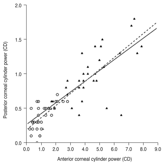
Fig. 1 Linear regression analysis of the relationship between anterior and posterior corneal cylinder power. The solid line represents a linear regression line (Y = 0.156X + 0.270, R2 = 0.470, p < 0.001) for the keratoconus group (filled triangles), and the dashed line represents a linear regression line (Y = 0.174X + 0.194, R2 = 0.422, p < 0.001) for the normal controls (open circles). CD = cylinder diopters.
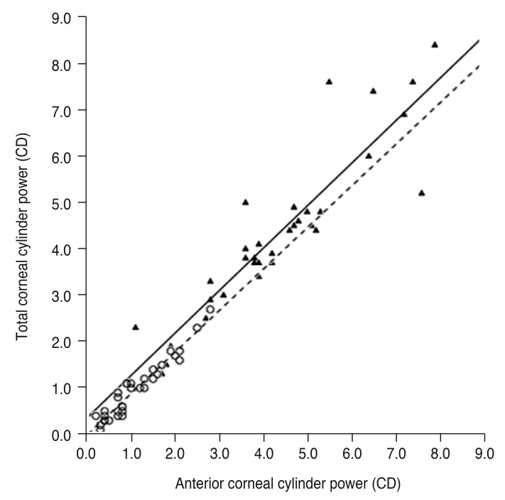
Fig. 2 Linear regression analysis of the relationship between anterior and total corneal cylinder power. The solid line represents a linear regression line (Y = 0.914X + 0.361, R2 = 0.824, p < 0.001) for the keratoconus group (filled triangles), and the dashed line represents a linear regression line (Y = 0.894X − 0.011, R2 = 0.928, p < 0.001) for the normal controls (open circles). CD = cylinder diopters.
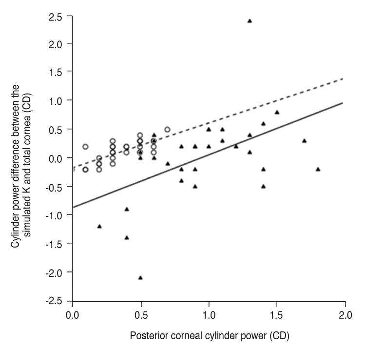
Fig. 3 Linear regression analysis of the relationship between posterior corneal cylinder power and cylinder power difference between the simulated keratometry (K) and total cornea. The solid line represents a linear regression line (Y = 0.928X − 0.868, R2 = 0.240, p = 0.004) for the keratoconus group (filled triangles), and the dashed line represents a linear regression line (Y = 0.786X − 0.172, R2 = 0.592, p < 0.001) for the normal controls (open circles). CD = cylinder diopters.
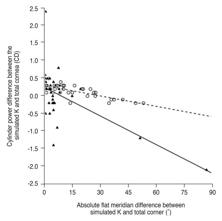
Fig. 4 Linear regression analysis of the relationship between absolute flat meridian difference and cylinder power difference between the simulated keratometry (K) and total cornea. The solid line represents a linear regression line (Y = −0.027X + 0.249, R2 = 0.370, p < 0.001) for the keratoconus group (filled triangles), and the dashed line represents a linear regression line (Y = −0.010X + 0.300, R2 = 0.592, p < 0.001) for the normal controls (open circles). CD = cylinder diopters.
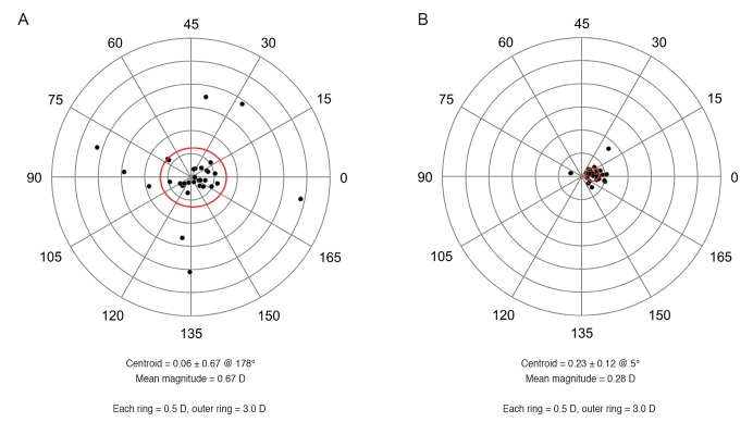
Fig. 5 Double-angle plots of the vector difference between the astigmatism of the anterior corneal surface and total cornea. The red ellipse indicates one standard deviation. (A) Keratoconus group. (B) Normal controls. D = diopters.
Reference
-
2. Wittig-Silva C, Chan E, Islam FM, et al. A randomized, controlled trial of corneal collagen cross-linking in progressive keratoconus: three-year results. Ophthalmology. 2014; 121:812–821. PMID: 24393351.3. Guell JL, Morral M, Malecaze F, et al. Collagen crosslinking and toric iris-claw phakic intraocular lens for myopic astigmatism in progressive mild to moderate keratoconus. J Cataract Refract Surg. 2012; 38:475–484. PMID: 22261324.4. Eom Y, Nam KT, Kang SY, et al. Axis difference between corneal and internal astigmatism to consider for toric intraocular lenses. Am J Ophthalmol. 2013; 156:1112–1119. PMID: 23972893.
Article5. Koch DD, Ali SF, Weikert MP, et al. Contribution of posterior corneal astigmatism to total corneal astigmatism. J Cataract Refract Surg. 2012; 38:2080–2087. PMID: 23069271.
Article6. Eom Y, Kang SY, Kim HM, Song JS. The effect of posterior corneal flat meridian and astigmatism amount on the total corneal astigmatism estimated f rom anterior corneal measurements. Graefes Arch Clin Exp Ophthalmol. 2014; 252:1769–1777. PMID: 25038911.7. Eom Y, Rhim JW, Kang SY, et al. Toric intraocular lens calculations using ratio of anterior to posterior corneal cylinder power. Am J Ophthalmol. 2015; 160:717–724. PMID: 26215437.
Article8. Eom Y, Ryu D, Kim DW, et al. Development of a program for toric intraocular lens calculation considering posterior corneal astigmatism, incision-induced posterior corneal astigmatism, and effective lens position. Graefes Arch Clin Exp Ophthalmol. 2016; 254:1977–1986. PMID: 27541160.
Article9. Tomidokoro A, Oshika T, Amano S, et al. Changes in anterior and posterior corneal curvatures in keratoconus. Ophthalmology. 2000; 107:1328–1332. PMID: 10889107.
Article10. Pinero DP, Alio JL, Aleson A, et al. Corneal volume, pachymetry, and correlation of anterior and posterior corneal shape in subclinical and different stages of clinical keratoconus. J Cataract Refract Surg. 2010; 36:814–825. PMID: 20457375.11. Montalban R, Alio JL, Javaloy J, Pinero DP. Correlation of anterior and posterior corneal shape in keratoconus. Cornea. 2013; 32:916–921. PMID: 23665644.12. Alio JL, Pena-Garcia P, Abdulla Guliyeva F, et al. MICS with toric intraocular lenses in keratoconus: outcomes and predictability analysis of postoperative refraction. Br J Ophthalmol. 2014; 98:365–370. PMID: 24390170.
Article13. Muftuoglu O, Ayar O, Ozulken K, et al. Posterior c orneal elevation and back difference corneal elevation in diagnosing forme fruste keratoconus in the fellow eyes of unilateral keratoconus patients. J Cataract Refract Surg. 2013; 39:1348–1357. PMID: 23820305.14. Krumeich JH, Daniel J, Knulle A. Live-epikeratophakia for keratoconus. J Cataract Refract Surg. 1998; 24:456–463. PMID: 9584238.
Article15. Choi JA, Kim MS. Progression of keratoconus by longitudinal assessment with corneal topography. Invest Ophthalmol Vis Sci. 2012; 53:927–935. PMID: 22247476.
Article16. Pinero DP, Camps VJ, Caravaca-Arens E, et al. Estimation of the central corneal power in keratoconus: theoretical and clinical assessment of the error of the keratometric approach. Cornea. 2014; 33:274–279. PMID: 24452214.17. Holladay JT, Moran JR, Kezirian GM. Analysis of aggregate surgically induced refractive change, prediction error, and intraocular astigmatism. J Cataract Refract Surg. 2001; 27:61–79. PMID: 11165858.
Article18. Savini G, Naeser K, Schiano-Lomoriello D, Mularoni A. Influence of posterior corneal astigmatism on total corneal astigmatism in eyes with keratoconus. Cornea. 2016; 35:1427–1433. PMID: 27387567.
Article19. Fam HB, Lim KL. Validity of the keratometric index: large population-based study. J Cataract Refract Surg. 2007; 33:686–691. PMID: 17397744.
Article20. Olsen T. On the calculation of power from curvature of the cornea. Br J Ophthalmol. 1986; 70:152–154. PMID: 3947615.
Article21. Ho JD, Tsai CY, Tsai RJ, et al. Validity of the keratometric index: evaluation by the Pentacam rotating Scheimpflug camera. J Cataract Refract Surg. 2008; 34:137–145. PMID: 18165094.
Article
- Full Text Links
-
- Actions
-
Cited
- CITED
-
- Close
- Share
-
- Similar articles
-
- Comparing Changes in Corneal Astigmatism Using Scheimpflug Camera after Epiblepharon Correction Surgery
- Comparison of Anterior and Posterior Elevation, and Sagittal Curvature between Keratoconus and Normal Cornea
- The Change in Corneal Astigmatism after Cataract Surgery in Patients with Small Amount of Astigmatism
- Corneal Topographic Study Using Orbscan II between Keratoconus and Keratoconus Suspect
- Corneal Thickness Measured by Dual Scheimpflug, Anterior Segment Optical Coherence Tomography, and Ultrasound Pachymetry
