Flexible Intramedullary Nailing of Forearm Fractures at the Distal Metadiaphyseal Junction in Adolescents
- Affiliations
-
- 1Department of Orthopedic Surgery, Soonchunhyang University Bucheon Hospital, Bucheon, Korea. reinhart98@hanmail.net
- KMID: 2412311
- DOI: http://doi.org/10.4055/cios.2017.9.1.101
Abstract
- BACKGROUND
The purpose of this study was to analyze the radiographic and functional outcomes of flexible intramedullary (IM) nailing in adolescent patients with forearm fractures at the diaphysis or at the metadiaphyseal junction (MDJ).
METHODS
We retrospectively reviewed the results of 40 patients who underwent IM nailing for pediatric forearm fractures. Thirty males and 10 females were followed for an average of 16 months (range, 12 to 20 months). Their average age was 11 years (range, 10 to 16 years). The average duration from the onset of trauma to surgery was 3.8 days (range, 1 to 36 days). Fracture sites were located at the MDJ of the radius in 8 patients (MDJ group) while 32 patients had middle-third fractures (D group). We assessed the magnitude and location of the maximum radial bow and range of movements. Functional outcomes were evaluated using Daruwalla criteria.
RESULTS
Open reduction was carried out in 8 cases. Union was achieved at an average of 8.3 weeks postoperatively. The results were classified as good in 38 and excellent in 2 according to Daruwalla criteria with restoration of forearm rotation. The mean angulation at the last follow-up was 1.8° on the anteroposterior radiograph and 3.3° on the lateral radiograph (MDJ group: 1.8° and 2.1°, respectively; D group: 1.9° and 2.8°, respectively). There was no significant difference in the mean angulation between the groups. The mean magnitude of maximal radial bow was 5.7% ± 1.8% (MDJ group, 5.2% ± 0.8%; D group, 5.9% ± 1.9%). The mean location of maximal radial bow was 58.0% ± 8.8% (MDJ group, 56.4% ± 8.9%; D group, 58.6% ± 8.9%). The differences in the mean magnitude and location of maximal radial bow with the normal contralateral arms (7.0% ± 1.2% and 50.9% ± 6.0%, respectively) were not significantly different between the groups. Complications included superficial infection (2), delayed union (1), and refracture (1).
CONCLUSIONS
IM nail fixation provided satisfactory results and maintained adequate stability for both forearm bone fractures in adolescents, even though the fracture was located at the MDJ of the radius.
MeSH Terms
-
Adolescent
Child
Diaphyses
Female
Forearm/physiopathology
Fracture Fixation, Intramedullary/adverse effects/*methods
*Fracture Healing
Humans
Male
Open Fracture Reduction
Radiography
Radius/*diagnostic imaging
Radius Fractures/diagnostic imaging/physiopathology/*surgery
Retrospective Studies
Rotation
Ulna Fractures/diagnostic imaging/physiopathology/*surgery
Figure
-
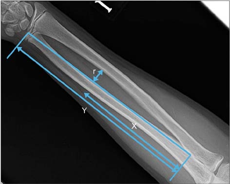
Fig. 1 Measurement methods of the magnitude and location of the maximum radial bow expressed as a percentage of the radial length. Magnitude of radial bow = (r/Y) × 100. Location of radial bow = (X/Y) × 100.
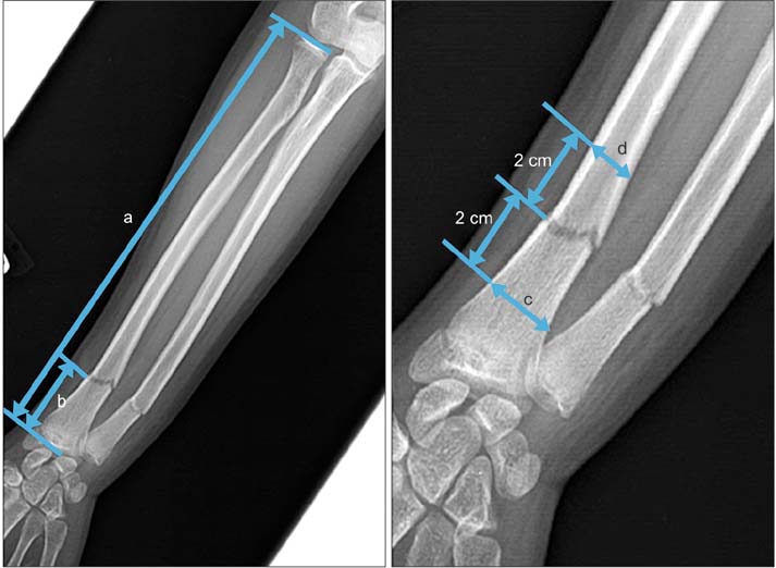
Fig. 2 Fracture at the metadiaphyseal junction (MDJ) was defined as a fracture with: (1) the distance (b) between the fracture line and the distal articular surface between 35 mm and 60 mm; (2) the ratio of the length of distal fragment (b) to the total length of radius (a) within 25%; and (3) the ratio of the maximal diameter at 2 cm proximal to the fracture line (d) over the maximal diameter (c) at 2 cm distal to the fracture line within 70%.

Fig. 3 (A) Radiograph of a 13-year-old boy (case 6) who had both forearm bone fractures at the metadiaphyseal junction. (B) Anteroposterior (left) and lateral (right) radiographs revealing intramedullary fixation of both radius and ulna. (C) Final follow-up radiographs showing bony union.
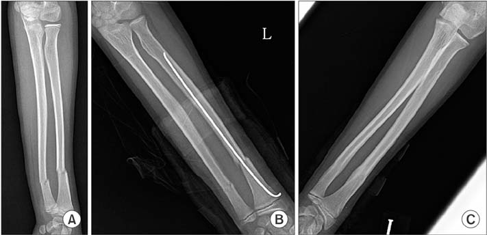
Fig. 4 (A) Radiograph of a 12-year-old boy (case 7) who had both forearm bone fractures at the metadiaphyseal junction. (B) Radiograph revealing intramedullary fixation of the radius. (C) Final follow-up radiograph showing bony union.
Cited by 1 articles
-
Intramedullary Blunt K-Wire Fixation for Pediatric Forearm Fractures
Yoo Joon Sur, Yong Sin Cho, Ho Youn Park
Arch Hand Microsurg. 2019;24(2):126-132. doi: 10.12790/ahm.2019.24.2.126.
Reference
-
1. Kapoor V, Theruvil B, Edwards SE, Taylor GR, Clarke NM, Uglow MG. Flexible intramedullary nailing of displaced diaphyseal forearm fractures in children. Injury. 2005; 36(10):1221–1225.
Article2. Reinhardt KR, Feldman DS, Green DW, Sala DA, Widmann RF, Scher DM. Comparison of intramedullary nailing to plating for both-bone forearm fractures in older children. J Pediatr Orthop. 2008; 28(4):403–409.
Article3. Rodriguez-Merchan EC. Pediatric fractures of the forearm. Clin Orthop Relat Res. 2005; (432):65–72.4. Bae DS. Pediatric distal radius and forearm fractures. J Hand Surg Am. 2008; 33(10):1911–1923.
Article5. Firl M, Wunsch L. Measurement of bowing of the radius. J Bone Joint Surg Br. 2004; 86(7):1047–1049.
Article6. Daruwalla JS. A study of radioulnar movements following fractures of the forearm in children. Clin Orthop Relat Res. 1979; (139):114–120.
Article7. Lascombes P, Prevot J, Ligier JN, Metaizeau JP, Poncelet T. Elastic stable intramedullary nailing in forearm shaft fractures in children: 85 cases. J Pediatr Orthop. 1990; 10(2):167–171.
Article8. Shah AS, Lesniak BP, Wolter TD, Caird MS, Farley FA, Vander Have KL. Stabilization of adolescent both-bone forearm fractures: a comparison of intramedullary nailing versus open reduction and internal fixation. J Orthop Trauma. 2010; 24(7):440–447.
Article9. Yung PS, Lam CY, Ng BK, Lam TP, Cheng JC. Percutaneous transphyseal intramedullary Kirschner wire pinning: a safe and effective procedure for treatment of displaced diaphyseal forearm fracture in children. J Pediatr Orthop. 2004; 24(1):7–12.
Article10. Battle J, Carmichael KD, Morris RP. Biomechanical comparison of flexible intramedullary nailing versus crossed Kirschner wire fixation in a canine model of pediatric forearm fractures. J Pediatr Orthop B. 2006; 15(5):370–375.
Article11. Dietz JF, Bae DS, Reiff E, Zurakowski D, Waters PM. Single bone intramedullary fixation of the ulna in pediatric both bone forearm fractures: analysis of short-term clinical and radiographic results. J Pediatr Orthop. 2010; 30(5):420–424.
Article12. Westacott D, Dickenson E, Smith N. Single-bone fixation of paediatric diaphyseal both-bone forearm fractures: a systematic review. Acta Orthop Belg. 2012; 78(4):425–430.
- Full Text Links
-
- Actions
-
Cited
- CITED
-
- Close
- Share
-
- Similar articles
-
- Operative Treatment in Fractures of the Humeral Shaft: Comparison of the Clinical Results of Flexible Intramedullary Nailing Versus Interlocking Intramedullary Nailing
- Comparison of Flexible Intramedullary Nailing with External Fixation for Treating Pediatric Femoral Shaft Fractures
- Interlocking Intramedullary Nailing Versus conventional Kuntscher Intramedullary Nailing for Fracture of the Femoral Shaft
- Flexible Intramedullary Nailing for Femoral Fractures
- Pediatric Forearm Bone Fractures Treated with Flexible Intramedullary Nail
