Ultrasonographic and Clinical Characteristics of Schwannoma of the Hand
- Affiliations
-
- 1Department of Orthopedic Surgery, Konkuk University School of Medicine, Seoul, Korea. lsjmd@kuh.ac.kr
- KMID: 2412309
- DOI: http://doi.org/10.4055/cios.2017.9.1.91
Abstract
- BACKGROUND
The purpose of this study was to report the ultrasonographic findings and clinical features of schwannoma of the hand.
METHODS
We enrolled 8 patients who were initially diagnosed with ganglion by ultrasonography but finally with schwannoma by a tissue biopsy. We retrospectively analyzed the ultrasonographic findings of eight patients including echogenicity, internal homogeneity, posterior enhancement, internal vascularity, and clinical manifestations such as the occurrence site, tenderness, Tinel's sign, and paresthesia before the surgery.
RESULTS
The occurrence sites were as follows: two cases on the thenar area, one case on the second web space, three cases on the third web space, one case on the radiovolar aspect of the proximal phalanx of the index finger, and one case on the radiovolar aspect of the proximal phalanx of the middle finger. Four patients suffered from tenderness and pain on presentation, and all patients had pain around the mass before presentation. Tinel's sign was present without paresthesia in one case. Ultrasonography revealed cystic lesions showing clear margins in all cases, and two of them had acoustic enhancement without internal flow.
CONCLUSIONS
It may not be easy to diagnosis schwannoma of the hand with ultrasonography alone when the lesion is small because of the similarity to the ultrasonographic findings of ganglion. Therefore, it is necessary to consider the possibility of schwannoma if a mass near the digital nerve or cutaneous nerve branch is accompanied by dull pain and tenderness.
Keyword
MeSH Terms
Figure
-
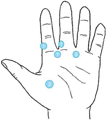
Fig. 1 The occurrence sites of schwannoma in the hand. The circled number indicates the number of occurrences.
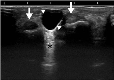
Fig. 2 Ultrasonographic findings. Axial scan shows homogenous, anechoic round mass (arrow head) with posterior enhancement (asterisk) and flexor tendon of finger (arrow).
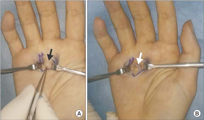
Fig. 3 Intraoperative photographs. (A) The yellowish-white colored ovoid mass (black arrow) was identified in the nerve at the bifurcation between the common digital nerve of the long and the ring fingers. (B) The yellowish-white colored solitary mass was enucleated microscopically. The white arrow indicates the digital nerve.
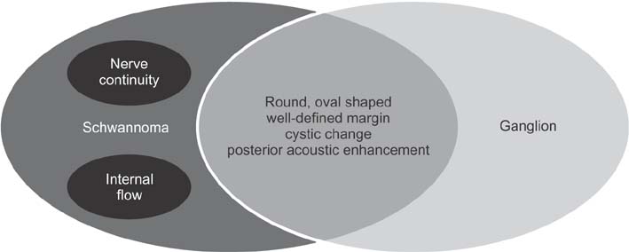
Fig. 4 Ultrasonographic features of ganglion and schwannoma. Schwannoma is often hypoechoic with posterior acoustic enhancement and thus may simulate a ganglion cyst. The diagnostic clue of schwannoma is nerve continuity.
Reference
-
1. Bianchi S, Abdelwahab IF, Zwass A, Giacomello P. Ultrasonographic evaluation of wrist ganglia. Skeletal Radiol. 1994; 23(3):201–203.
Article2. Kransdorf MJ, Murphey MD. Imaging of soft tissue tumors. Philadelphia, PA: W.B. Saunders;1997. p. 275–316.3. Das Gupta TK, Brasfield RD, Strong EW, Hajdu SI. Benign solitary Schwannomas (neurilemomas). Cancer. 1969; 24(2):355–366.
Article4. Rockwell GM, Thoma A, Salama S. Schwannoma of the hand and wrist. Plast Reconstr Surg. 2003; 111(3):1227–1232.
Article5. White NB. Neurilemomas of the extremities. J Bone Joint Surg Am. 1967; 49(8):1605–1610.
Article6. Simoens WA, Wuyts FL, De Beuckeleer LH, Vandevenne JE, Bloem JL, De Schepper AM. MR features of peripheral nerve sheath tumors: can a calculated index compete with radiologist's experience? Eur Radiol. 2001; 11(2):250–257.
Article7. Reynolds DL Jr, Jacobson JA, Inampudi P, Jamadar DA, Ebrahim FS, Hayes CW. Sonographic characteristics of peripheral nerve sheath tumors. AJR Am J Roentgenol. 2004; 182(3):741–744.
Article8. Lin J, Jacobson JA, Hayes CW. Sonographic target sign in neurofibromas. J Ultrasound Med. 1999; 18(7):513–517.
Article9. Chinn DH, Filly RA, Callen PW. Unusual ultrasonographic appearance of a solid schwannoma. J Clin Ultrasound. 1982; 10(5):243–245.
Article10. Hughes DG, Wilson DJ. Ultrasound appearances of peripheral nerve tumours. Br J Radiol. 1986; 59(706):1041–1043.
Article11. Murphey MD, Smith WS, Smith SE, Kransdorf MJ, Temple HT. From the archives of the AFIP: imaging of musculoskeletal neurogenic tumors: radiologic-pathologic correlation. Radiographics. 1999; 19(5):1253–1280.12. Bacigalupo L, Bianchi S, Valle M, Martinoli C. Ultrasonography of peripheral nerves. Radiologe. 2003; 43(10):841–849.13. Stuart RM, Koh ES, Breidahl WH. Sonography of peripheral nerve pathology. AJR Am J Roentgenol. 2004; 182(1):123–129.
Article14. Kececi Y, Gurler T, Gundogan H, Bilkay U, Cagdas A. Benign giant schwannoma located in the upper arm. Ann Plast Surg. 1997; 39(1):100–102.
Article15. Kang HJ, Shin SJ, Kang ES. Schwannomas of the upper extremity. J Hand Surg Br. 2000; 25(6):604–607.
Article16. De Flaviis L, Nessi R, Del Bo P, Calori G, Balconi G. High-resolution ultrasonography of wrist ganglia. J Clin Ultrasound. 1987; 15(1):17–22.
Article17. Kehoe NJ, Reid RP, Semple JC. Solitary benign peripheral-nerve tumours: review of 32 years' experience. J Bone Joint Surg Br. 1995; 77(3):497–500.
Article18. Holdsworth BJ. Nerve tumours in the upper limb: a clinical review. J Hand Surg Br. 1985; 10(2):236–238.
Article19. Tsai WC, Chiou HJ, Chou YH, Wang HK, Chiou SY, Chang CY. Differentiation between schwannomas and neurofibromas in the extremities and superficial body: the role of high-resolution and color Doppler ultrasonography. J Ultrasound Med. 2008; 27(2):161–166.
Article20. Plate AM, Lee SJ, Steiner G, Posner MA. Tumorlike lesions and benign tumors of the hand and wrist. J Am Acad Orthop Surg. 2003; 11(2):129–141.
Article21. Carra BJ, Bui-Mansfield LT, O'Brien SD, Chen DC. Sonography of musculoskeletal soft-tissue masses: techniques, pearls, and pitfalls. AJR Am J Roentgenol. 2014; 202(6):1281–1290.
Article22. Peer S, Kovacs P, Harpf C, Bodner G. High-resolution sonography of lower extremity peripheral nerves: anatomic correlation and spectrum of disease. J Ultrasound Med. 2002; 21(3):315–322.23. Rinaldi E. Neurilemomas and neurofibromas of the upper limb. J Hand Surg Am. 1983; 8(5 Pt 1):590–593.
Article
- Full Text Links
-
- Actions
-
Cited
- CITED
-
- Close
- Share
-
- Similar articles
-
- Tibial Schwannoma Mimicking a Popliteal Cyst
- Atypically Located Brainstem Schwannoma
- Surgical treatment of multiple plexiform schwannomas arising from the superficial radial nerve: a case report
- Huge Plexiform Schwannoma of the Ulnar Nerve
- Subungual Schwannoma Mimicking Glomus Tumor on Ultrasonography: A Case Report
