The Validation of Ultrasound-Guided Target Segment Identification in Thoracic Spine as Confirmed by Fluoroscopy
- Affiliations
-
- 1Department of Orthopedic Surgery, Kwangju Christian Hospital, Gwangju, Korea. stemcellchoi@gmail.com
- KMID: 2412248
- DOI: http://doi.org/10.4055/cios.2017.9.4.472
Abstract
- BACKGROUND
The role of ultrasound in the thoracic spine has been underappreciated, partly because of the relative efficacy of the landmark-guided technique and the limitation of imaging through the narrow acoustic windows produced by the bony framework of thoracic spine. The aim of this study was to make a comparison between the 12th rib and the spinous process of C7 as a landmark for effective ultrasound-guided target segment identification in the thoracic spine.
METHODS
Ultrasonography of 44 thoracic spines was performed and the same procedure was carried out 1 week later again. The target segments (T3-4, T7-8, and T10-11) were identified using the 12th rib (group 1) or the spinous process of C7 (group 2) as a starting landmark. Ultrasound scanning was done proximally (group 1) or distally (group 2) toward the target transverse process and further medially and slightly superior to the target thoracic facet. Then, a metal marker was placed on the T3-4, T7-8, and T10-11 and the location of each marker was confirmed by fluoroscopy.
RESULTS
In the total 132 segments, sonographic identification was confirmed to be successful with fluoroscopy in 84.1% in group 1 and 56.8% in group 2. Group 1 had a greater success rate in ultrasound-guided target segment identification than group 2 (p = 0.001), especially in T10-11 (group 1, 93.2%; group 2, 43.2%; p = 0.001) and T7-8 (group 1, 86.4%; group 2, 56.8%; p = 0.002). The intrarater reliability of ultrasound-guided target segment identification was good (group 1, r = 0.76; group 2, r = 0.82), showing no difference between right and left sides. Ultrasound-guided target segment identification was more effective in the non-obese subjects (p = 0.001), especially in group 1.
CONCLUSIONS
Ultrasound-guided detection using the 12th rib as a starting landmark for scanning could be a promising technique for successful target segment identification in the thoracic spine.
Keyword
MeSH Terms
Figure
-
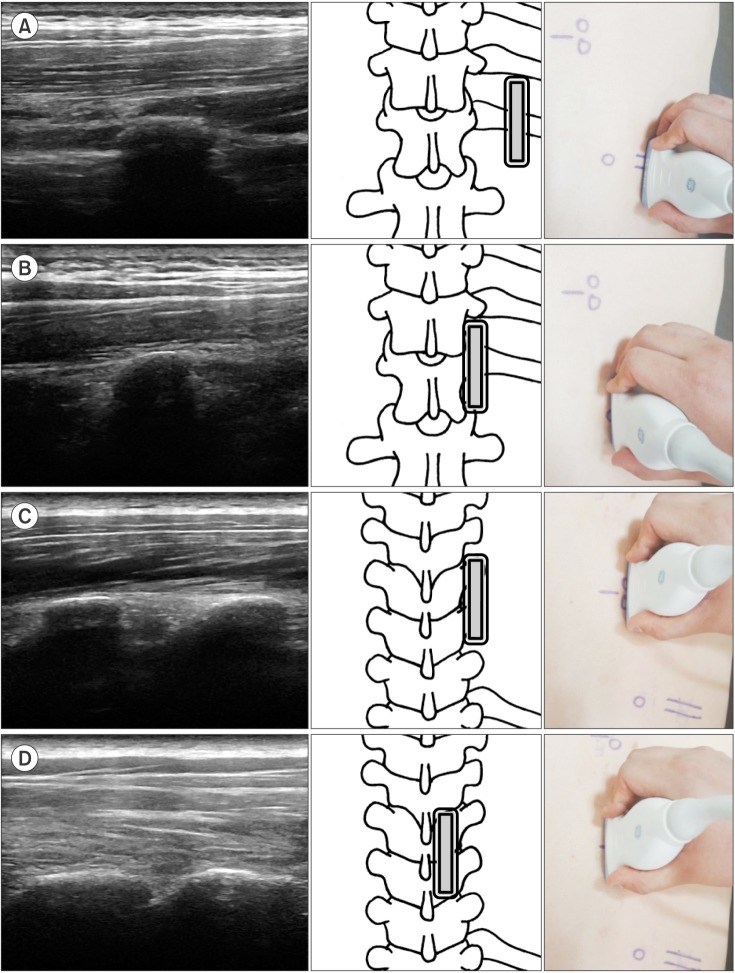
Fig. 1 Ultrasound images of the detection method using the 12th rib as a starting landmark. The double-lined rectangle represents the ultrasound probe. (A) The 12th rib in the sagittal view. (B) The transverse process of T12 was found medial to the 12th rib. (C) The probe was moved proximally to the transverse process of T7 and T8. (D) The facet joint of T7–8 was found medially and slightly proximal to the transverse process.
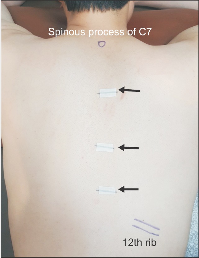
Fig. 2 Metal makers (black arrows) placed at the target location identified with sonography.
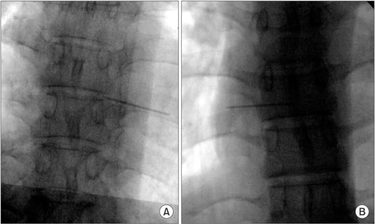
Fig. 3 Validation by C-arm fluoroscopy. (A) Successful case: the metal marker was accurately located at the facet joint of the target segment. (B) Failure case: the metal marker was not accurately located at the facet joint of the target segment.
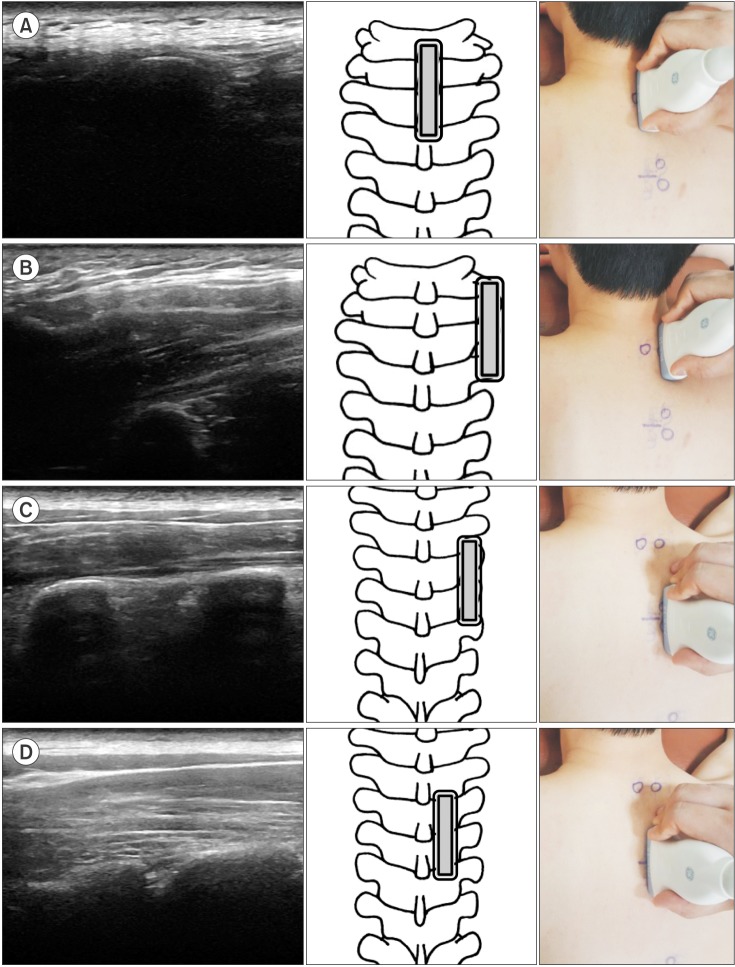
Fig. 4 Ultrasound images of the detection method using the spinous process of C7 as a starting landmark. The double-lined rectangle represents the ultrasound probe. (A) The spinous process of C7 in the sagittal view. (B) The transverse process of T1 was found lateral to the spinous process of C7. (C) The probe was moved distally to the transverse process of T3 and T4. (D) The facet joint of T3–4 was found medially and slight proximal to the transverse process.
Reference
-
1. Linton SJ, Hellsing AL, Hallden K. A population-based study of spinal pain among 35-45-year-old individuals: prevalence, sick leave, and health care use. Spine (Phila Pa 1976). 1998; 23(13):1457–1463. PMID: 9670397.2. Leboeuf-Yde C, Nielsen J, Kyvik KO, Fejer R, Hartvigsen J. Pain in the lumbar, thoracic or cervical regions do age and gender matter A population-based study of 34,902 Danish twins 20-71 years of age. BMC Musculoskelet Disord. 2009; 10:39. PMID: 19379477.
Article3. Schnabel A, Reichl SU, Kranke P, Pogatzki-Zahn EM, Zahn PK. Efficacy and safety of paravertebral blocks in breast surgery: a meta-analysis of randomized controlled trials. Br J Anaesth. 2010; 105(6):842–852. PMID: 20947592.
Article4. Manchikanti L, Singh V, Falco FJ, Cash KA, Pampati V, Fellows B. Comparative effectiveness of a one-year follow-up of thoracic medial branch blocks in management of chronic thoracic pain: a randomized, double-blind active controlled trial. Pain Physician. 2010; 13(6):535–548. PMID: 21102966.5. Park KD, Jee H, Nam HS, et al. Effect of medial branch block in chronic facet joint pain for osteoporotic compression fracture: one year retrospective study. Ann Rehabil Med. 2013; 37(2):191–201. PMID: 23705113.
Article6. Mitra R, Do H, Alamin T, Cheng I. Facet pain in thoracic compression fractures. Pain Med. 2010; 11(11):1674–1677. PMID: 21029349.
Article7. Moon SH, Lee S, Lee JI. Ultrasound-guided intervention in thoracic spine. J Korean Orthop Assoc. 2015; 50(2):93–106.
Article8. Stulc SM, Hurdle MF, Pingree MJ, Brault JS, Porter CA. Ultrasound-guided thoracic facet injections: description of a technique. J Ultrasound Med. 2011; 30(3):357–362. PMID: 21357557.9. O Riain SC, Donnell BO, Cuffe T, Harmon DC, Fraher JP, Shorten G. Thoracic paravertebral block using real-time ultrasound guidance. Anesth Analg. 2010; 110(1):248–251. PMID: 19933536.10. Deimel GW, Hurdle MF, Murthy N, Cartwright JA, Smith J, Pingree MJ. Sonographically guided costotransverse joint injections: a computed tomographically controlled cadaveric feasibility study. J Ultrasound Med. 2013; 32(12):2083–2089. PMID: 24277889.11. Marhofer P, Kettner SC, Hajbok L, Dubsky P, Fleischmann E. Lateral ultrasound-guided paravertebral blockade: an anatomical-based description of a new technique. Br J Anaesth. 2010; 105(4):526–532. PMID: 20685684.
Article12. Choi DH, Jung HG, Lee JH, Park JH, Choi YS. Effectiveness of Doppler image of the vertebral artery as an anatomical landmark for identification of ultrasound-guided target level in cervical spine. Asian Spine J. 2015; 9(5):683–688. PMID: 26435784.
Article13. Narouze SN, Vydyanathan A, Kapural L, Sessler DI, Mekhail N. Ultrasound-guided cervical selective nerve root block: a fluoroscopy-controlled feasibility study. Reg Anesth Pain Med. 2009; 34(4):343–348. PMID: 19574867.14. Greher M, Scharbert G, Kamolz LP, et al. Ultrasound-guided lumbar facet nerve block: a sonoanatomic study of a new methodologic approach. Anesthesiology. 2004; 100(5):1242–1248. PMID: 15114223.15. Galiano K, Obwegeser AA, Bodner G, et al. Ultrasound-guided periradicular injections in the middle to lower cervical spine: an imaging study of a new approach. Reg Anesth Pain Med. 2005; 30(4):391–396. PMID: 16032592.
Article16. Mehta MH, Patel RV, Mehta LV, Bhatt YC. Congenital absence of ribs. Indian Pediatr. 1992; 29(9):1149–1152. PMID: 1452315.17. Saranteas T. Limitations in ultrasound imaging techniques in anesthesia: obesity and muscle atrophy? Anesth Analg. 2009; 109(3):993–994. PMID: 19690282.
Article18. Rauch S, Kasuya Y, Turan A, Neamtu A, Vinayakan A, Sessler DI. Ultrasound-guided lumbar medial branch block in obese patients: a fluoroscopically confirmed clinical feasibility study. Reg Anesth Pain Med. 2009; 34(4):340–342. PMID: 19585701.19. Ueshima H, Kubo K, Sakamoto S, et al. A case of the transversus abdominis plane block in a super obese patient using a convex probe. Masui. 2013; 62(4):439–441. PMID: 23697197.
- Full Text Links
-
- Actions
-
Cited
- CITED
-
- Close
- Share
-
- Similar articles
-
- Ultrasound-Guided Intervention in Thoracic Spine
- The Validation of Ultrasound-Guided Lumbar Facet Nerve Blocks as Confirmed by Fluoroscopy
- Ultrasound-guided interventions for spinal pain
- Ultrasound-Guided Intervention in Lumbar Spine
- Feasibility of Ultrasound-Guided Lumbar and S1 Nerve Root Block: A Cadaver Study
