Clin Orthop Surg.
2017 Dec;9(4):405-412. 10.4055/cios.2017.9.4.405.
Factors Associated with the Risk of Articular Surface Perforation during Anchor Placement for Arthroscopic Acetabular Labral Repair
- Affiliations
-
- 1Department of Orthopaedic Surgery, Gyeongsang National University Hospital, Gyeongsang National University School of Medicine, Jinju, Korea.
- 2Department of Orthopaedic Surgery, Chung-Ang University College of Medicine, Seoul, Korea. hayongch@naver.com
- 3Department of Orthopedic Surgery, Seoul National University Bundang Hospital, Seongnam, Korea.
- KMID: 2412240
- DOI: http://doi.org/10.4055/cios.2017.9.4.405
Abstract
- BACKGROUND
The purpose of this study was to evaluate factors associated with the risk of articular surface perforation during anchor placement for arthroscopic acetabular labral repair using follow-up computed tomographic arthrography (CTA).
METHODS
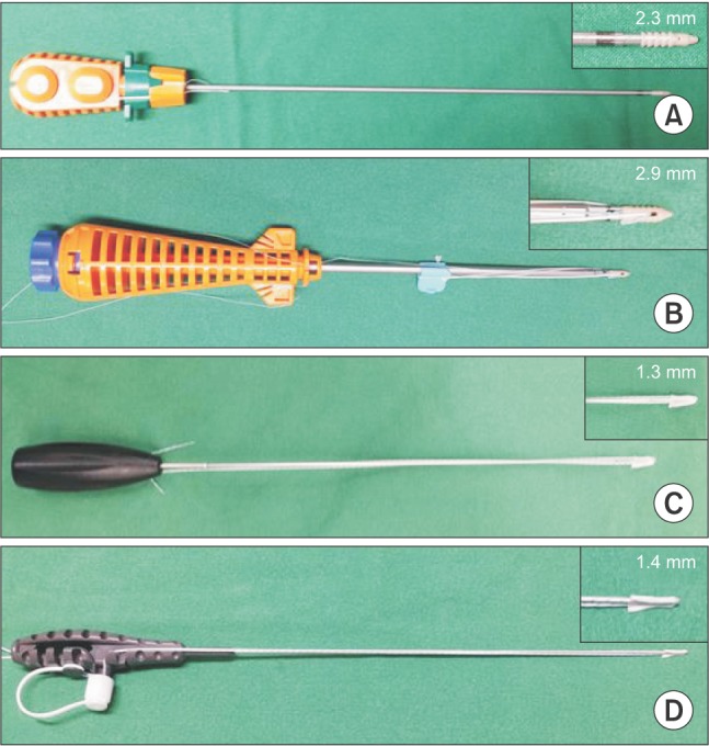
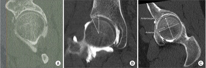
Forty-six patients (29 males and 17 females) underwent arthroscopic labral repair using 142 suture anchors (55 large anchors and 87 small anchors). The patients were followed with CTA 1 year postoperatively. Anchor position was assessed by the insertion angle and the distance of the suture anchor tip from the articular cartilage. The incidence of malposition of suture anchors was assessed in follow-up CTA. The location and incidence of malposition were compared between two groups divided according to the diameter of suture anchor.
RESULTS
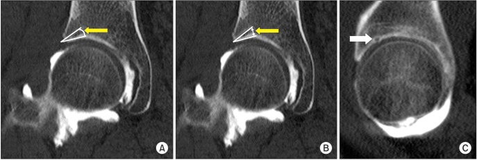
The mean insertion angle and distance were significantly different between the groups. Of the 142 anchors, 15 (11%) were placed in the cartilage-bone transitional zone. Articular involvement was most common at the 3 o'clock position of the suture anchor (six out of 33 anchors, 18.2%). Both the insertion angle and distance showed small values in the articular involvement group.
CONCLUSIONS
The radiographic analysis of the placement of suture anchors after arthroscopic labral refixation based on follow-up CTA demonstrates that articular involvement of anchors is related to the location on the acetabular rim (clock position) and anchor diameter.
Keyword
MeSH Terms
-
Acetabulum/diagnostic imaging/*surgery
Adult
Arthrography
Arthroscopy/adverse effects
Cartilage, Articular/diagnostic imaging/*injuries
Female
Hip Joint/diagnostic imaging/*surgery
Humans
Male
Middle Aged
Prosthesis Implantation/*adverse effects
Retrospective Studies
Risk Factors
Suture Anchors/*adverse effects
Tomography, X-Ray Computed
Young Adult
Figure
Reference
-
1. Haddad B, Konan S, Haddad FS. Debridement versus re-attachment of acetabular labral tears: a review of the literature and quantitative analysis. Bone Joint J. 2014; 96-B(1):24–30. PMID: 24395306.2. Krych AJ, Kuzma SA, Kovachevich R, Hudgens JL, Stuart MJ, Levy BA. Modest mid-term outcomes after isolated arthroscopic debridement of acetabular labral tears. Knee Surg Sports Traumatol Arthrosc. 2014; 22(4):763–767. PMID: 24493256.
Article3. Larson CM, Giveans MR. Arthroscopic debridement versus refixation of the acetabular labrum associated with femoroacetabular impingement. Arthroscopy. 2009; 25(4):369–376. PMID: 19341923.
Article4. Philippon MJ, Briggs KK, Yen YM, Kuppersmith DA. Outcomes following hip arthroscopy for femoroacetabular impingement with associated chondrolabral dysfunction: minimum two-year follow-up. J Bone Joint Surg Br. 2009; 91(1):16–23. PMID: 19091999.5. Crawford MJ, Dy CJ, Alexander JW, et al. The 2007 Frank Stinchfield Award: the biomechanics of the hip labrum and the stability of the hip. Clin Orthop Relat Res. 2007; 465:16–22. PMID: 17906586.
Article6. Ferguson SJ, Bryant JT, Ganz R, Ito K. An in vitro investigation of the acetabular labral seal in hip joint mechanics. J Biomech. 2003; 36(2):171–178. PMID: 12547354.
Article7. Jackson TJ, Hammarstedt JE, Vemula SP, Domb BG. Acetabular labral base repair versus circumferential suture repair: a matched-paired comparison of clinical outcomes. Arthroscopy. 2015; 31(9):1716–1721. PMID: 25911393.
Article8. Kelly BT, Weiland DE, Schenker ML, Philippon MJ. Arthroscopic labral repair in the hip: surgical technique and review of the literature. Arthroscopy. 2005; 21(12):1496–1504. PMID: 16376242.
Article9. Philippon MJ, Schroder e Souza BG, Briggs KK. Labrum: resection, repair and reconstruction sports medicine and arthroscopy review. Sports Med Arthrosc. 2010; 18(2):76–82. PMID: 20473125.
Article10. Matsuda DK, Bharam S, White BJ, Matsuda NA, Safran M. Anchor-induced chondral damage in the hip. J Hip Preserv Surg. 2015; 2(1):56–64. PMID: 27011815.
Article11. Hernandez JD, McGrath BE. Safe angle for suture anchor insertion during acetabular labral repair. Arthroscopy. 2008; 24(12):1390–1394. PMID: 19038710.
Article12. Lertwanich P, Ejnisman L, Torry MR, Giphart JE, Philippon MJ. Defining a safety margin for labral suture anchor insertion using the acetabular rim angle. Am J Sports Med. 2011; 39(Suppl):111S–116S. PMID: 21709040.
Article13. Nho SJ, Freedman RL, Federer AE, et al. Computed tomographic analysis of curved and straight guides for placement of suture anchors for acetabular labral refixation. Arthroscopy. 2013; 29(10):1623–1627. PMID: 24075612.
Article14. Thomas Byrd JW. Modified anterior portal for hip arthroscopy. Arthrosc Tech. 2013; 2(4):e337–e339. PMID: 24400178.
Article15. Philippon MJ, Stubbs AJ, Schenker ML, Maxwell RB, Ganz R, Leunig M. Arthroscopic management of femoroacetabular impingement: osteoplasty technique and literature review. Am J Sports Med. 2007; 35(9):1571–1580. PMID: 17420508.16. Landis JR, Koch GG. The measurement of observer agreement for categorical data. Biometrics. 1977; 33(1):159–174. PMID: 843571.
Article17. Foster AD, Ryan J, Ellis T, Flom J. Safe suture anchor insertion for anterior and posterior hip labral repair. J Hip Preserv Surg. 2015; 2(2):170–174. PMID: 27011835.
Article18. Stanton M, Banffy M. Safe angle of anchor insertion for labral repair during hip arthroscopy. Arthroscopy. 2016; 32(9):1793–1797. PMID: 27132777.
Article19. Ha YC, Choi JA, Lee YK, et al. The diagnostic value of direct CT arthrography using MDCT in the evaluation of acetabular labral tear: with arthroscopic correlation. Skeletal Radiol. 2013; 42(5):681–688. PMID: 23073899.
Article20. Jung JY, Kim GU, Lee HJ, Jang EC, Song IS, Ha YC. Diagnostic value of ultrasound and computed tomographic arthrography in diagnosing anterosuperior acetabular labral tears. Arthroscopy. 2013; 29(11):1769–1776. PMID: 24071389.
- Full Text Links
- Actions
-
Cited
- CITED
-
- Close
- Share
- Similar articles
-
- Arthroscopic Labral Repair Associated with Femoroacetabular Impingement: Short Term 2-5 Years Follow-up Results
- Treatment of Acetabular Avulsion Fracture with Labral Tear Using Suture Anchor: A Case Report
- Results According to Arthroscopic Repair Methods of Bankart Lesion (Operative Methods According to Labral Tear)
- Patient Satisfaction after Arthroscopic Repair of Acetabular Labral Tears
- Arthroscopic Management of Intraarticular Screw Perforation after Surgical Treatment of an Acetabular Posterior Wall Fracture: A Case Report


