Arthroscopic Management of Intraarticular Screw Perforation after Surgical Treatment of an Acetabular Posterior Wall Fracture: A Case Report
- Affiliations
-
- 1Department of Orthopaedic Surgery, Pusan National University Hospital, Busan, Korea. namhoonmoon@gmail.com
- KMID: 2408187
- DOI: http://doi.org/10.5371/hp.2018.30.1.60
Abstract
- Although surgical techniques for treating acetabular fracture are evolving, the use of periacetabular screws is common, and their placement in acetabular surgery is still technically demanding. For instance, intraarticular screw perforation is a serious complication that may occur during surgical treatment of an acetabular fracture. Here, we describe the case of a 50-year-old female who experienced an intraarticular screw perforation after surgical treatment of a posterior acetabular wall fracture. Removal of the perforated screw was performed arthroscopically based on its ability to offer minimally invasive access to the hip joint. One year after removal of the screw, no radiological signs of osteoarthritic changes were observed. The patient regained normal ambulation without limitations to range of motion or hip pain. To our knowledge, this is the first report on the use of arthroscopy to treat intraarticular screw perforation after surgical treatment of an acetabular fracture.
MeSH Terms
Figure
-
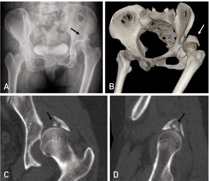
Fig. 1 (A, B) A 50-year-old woman with a left posterior acetabular wall fracture and posterior hip dislocation (black and white arrows). (C, D) Coronal and sagittal images of a computed tomography scan reveal a small osteochondral fragment (black arrow).
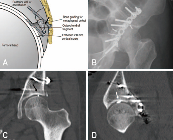
Fig. 2 (A) An embedded 2.0-mm cortical screw was used to fix the osteochondral fragment. (B) Postoperative radiographs show good congruency of the hip joint and well-fixed posterior wall fracture but reveal a possible intraarticular screw perforation (black arrow). (C, D) Coronal and sagittal images of a computed tomography scan confirm a screw perforation.
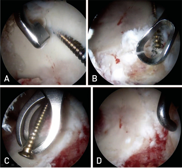
Fig. 3 (A) Arthroscopy showing intraarticular perforation of the cortical screw heading from 2 o'clock to 10 o'clock. (B) Articular cartilage surrounding the screw was removed using an extra small arthroscopic gauge and ring curette for whole exposure of the screw. (C) Arthroscopy showing basket forceps removing the lag screw. (D) Osteochondral defect and linear cartilage defect after screw removal.
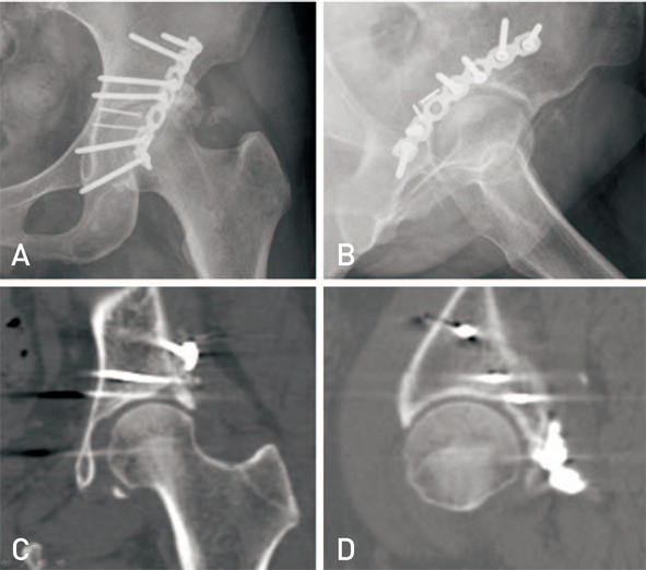
Fig. 4 No radiologic signs of osteoarthritic changes are observed, although a one year follow-up computed tomography revealed a small defect in the posterosuperior portion of the acetabulum.
Reference
-
1. Gras F, Marintschev I, Klos K, Mückley T, Hofmann GO, Kahler DM. Screw placement for acetabular fractures: which navigation modality (2-dimensional vs. 3-dimensional) should be used? An experimental study. J Orthop Trauma. 2012; 26:466–473.2. Eberl R, Müller EJ, Kaminski A, Muhr G. [The postoperative control CT after a fracture of the acetabulum. A useful quality control measure or an unnecessary exposure to radiation?]. Unfallchirurg. 2003; 106:741–745. German.3. Yamamoto Y, Ide T, Ono T, Hamada Y. Usefulness of arthroscopic surgery in hip trauma cases. Arthroscopy. 2003; 19:269–273.
Article4. Hari Krishnan B, Joshi GR, Pushkar A. Arthroscopic removal of intraarticular fracture fragment after fracture dislocation of hip. Med J Armed Forces India. 2015; 71:Suppl 1. S208–S210.
Article5. Ide T, Akamatsu N, Nakajima I. Arthroscopic surgery of the hip joint. Arthroscopy. 1991; 7:204–211.
Article6. Kim PS, Hwang DS. The current concepts of hip arthroscopy. J Korean Orthop Assoc. 2017; 52:484–499.
Article7. Park JY, Chung WC, Kim CK, Huh SH, Kim SJ, Jung BH. Arthroscopic reduction and transportal screw fixation of acetabular posterior wall fracture: technical note. Hip Pelvis. 2016; 28:120–126.
Article8. Enseki KR, Martin RL, Draovitch P, Kelly BT, Philippon MJ, Schenker ML. The hip joint: arthroscopic procedures and postoperative rehabilitation. J Orthop Sports Phys Ther. 2006; 36:516–525.
Article9. Kubo T, Utsunomiya H, Watanuki M, Hayashi H, Sakai A, Uchida S. Hip arthroscopic osteochondral autologous transplantation for treating osteochondritis dissecans of the femoral head. Arthrosc Tech. 2015; 4:e675–e680.
Article10. Fontana A, Bistolfi A, Crova M, Rosso F, Massazza G. Arthroscopic treatment of hip chondral defects: autologous chondrocyte transplantation versus simple debridement--a pilot study. Arthroscopy. 2012; 28:322–329.
Article
- Full Text Links
-
- Actions
-
Cited
- CITED
-
- Close
- Share
-
- Similar articles
-
- Arthroscopic Reduction and Transportal Screw Fixation of Acetabular Posterior Wall Fracture: Technical Note
- Surgical Treatment of Acetabular Posterior Wall Fracture with Hip Arthroscopy: A Case Report
- Single Percutaneous Retrograde Anterior Column Screw Fixation in a Minimally Displaced Transverse Acetabular Fracture - A Case Report -
- Treatment of the Acetabular Fracture
- Anatomical Results According to Fracture Pattern after Surgical Treatment of Acetabular Fractures
