Clin Orthop Surg.
2018 Jun;10(2):149-156. 10.4055/cios.2018.10.2.149.
Cortical Thickness Index of the Proximal Femur: A Radiographic Parameter for Preliminary Assessment of Bone Mineral Density and Osteoporosis Status in the Age 50 Years and Over Population
- Affiliations
-
- 1Department of Orthopaedic Surgery, Hamamatsu University School of Medicine, Hamamatsu, Japan. bao.ng@hama-med.ac.jp
- KMID: 2411739
- DOI: http://doi.org/10.4055/cios.2018.10.2.149
Abstract
- BACKGROUND
Bone mineral density (BMD) is the indicator of bone quality in at-risk individuals. Along with the fracture risk assessment tool (FRAX), a quick assessment of BMD from routine radiographs may be useful in the case of lacking X-ray absorptiometry data. This study aimed to investigate the correlation of cortical thickness index (CTI) and canal flare index (CFI) with BMD and FRAX and to evaluate their ability to predict femoral neck BMD (nBMD) and FRAX in the general elderly population.
METHODS
A total of 560 volunteers (age ≥ 50 years) who underwent hip-spine X-ray, BMD scanning and FRAX calculation were retrospectively reviewed. CTI and CFI were measured on anteroposterior radiographs and analyzed for their correlation with BMD and FRAX and for their ability to predict nBMD. The ability of CTI to predict osteoporosis status (OPS) and fracture risk status (FRS) was also investigated and the threshold values were calculated. All the analyses were performed separately on male and female subjects.
RESULTS
Significant differences in CTI, CFI, nBMD and FRAX between males and females were observed. CTI and CFI demonstrated significant positive correlation with nBMD and FRAX (all p < 0.001) in both males and females. CTI, height, and weight significantly predicted nBMD. CTI statistically predicted OPS and FRS, and the values of 0.56 and 0.62 were computed as CTI thresholds for males and females, respectively.
CONCLUSIONS
CTI was significantly correlated with nBMD and it predicted nBMD at good prediction levels. Therefore, CTI may be used as a supportive tool in the assessment of OPS and FRS besides BMD and FRAX in clinical practice.
MeSH Terms
Figure
-
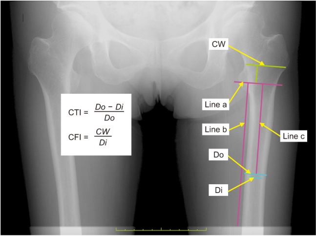
Fig. 1 Measurement of cortical thickness index (CTI) and canal flare index (CFI) on an anteroposterior radiograph (using Osirix software). Do: outer diameter (the shaft's outer diameter at 10 cm below the lesser trochanter), Di: inner diameter (the shaft's inner diameter at 10 cm below the lesser trochanter, measured at the same level as Do), CW: canal width (the canal width measured at 2 cm above Line a), Line a: a line drawn perpendicular to the femoral shaft through the middle point of the lesser trochanter, Line b: a line drawn parallel to the shaft to be used as a reference for drawing Line a, Line c: a 10-cm line drawn perpendicular to Line a, used to identify the shaft's inner and Do measurement levels.
Reference
-
1. NIH Consensus Development Panel on Osteoporosis Prevention, Diagnosis, and Therapy. Osteoporosis prevention, diagnosis, and therapy. JAMA. 2001; 285(6):785–795. PMID: 11176917.2. Masi L. Epidemiology of osteoporosis. Clin Cases Miner Bone Metab. 2008; 5(1):11–13. PMID: 22460840.3. Leboime A, Confavreux CB, Mehsen N, Paccou J, David C, Roux C. Osteoporosis and mortality. Joint Bone Spine. 2010; 77(Suppl 2):S107–S112. PMID: 21211746.
Article4. Hagino H, Sakamoto K, Harada A, et al. Nationwide one-decade survey of hip fractures in Japan. J Orthop Sci. 2010; 15(6):737–745. PMID: 21116890.
Article5. Kinoshita H, Tamaki T, Hashimoto T, Kasagi F. Factors influencing lumbar spine bone mineral density assessment by dual-energy X-ray absorptiometry: comparison with lumbar spinal radiogram. J Orthop Sci. 1998; 3(1):3–9. PMID: 9654549.
Article6. Mithal A, Ebeling P, Kyer CS. Asia-Pacific regional audit: epidemiology, costs and burden of osteoporosis in 2013 [Internet]. Nyon: International Osteoporosis Foundation;2013. cited 2018 Mar 9. Available from: https://www.iofbonehealth.org/datapublications/regional-audits/asia-pacific-regional-audit.7. Noble PC, Box GG, Kamaric E, Fink MJ, Alexander JW, Tullos HS. The effect of aging on the shape of the proximal femur. Clin Orthop Relat Res. 1995; (316):31–44. PMID: 7634721.
Article8. Tawada K, Iguchi H, Tanaka N, et al. Is the canal flare index a reliable means of estimation of canal shape? Measurement of proximal femoral geometry by use of 3D models of the femur. J Orthop Sci. 2015; 20(3):498–506. PMID: 25740729.
Article9. Dorr LD, Faugere MC, Mackel AM, Gruen TA, Bognar B, Malluche HH. Structural and cellular assessment of bone quality of proximal femur. Bone. 1993; 14(3):231–242. PMID: 8363862.
Article10. Noble PC, Alexander JW, Lindahl LJ, Yew DT, Granberry WM, Tullos HS. The anatomic basis of femoral component design. Clin Orthop Relat Res. 1988; (235):148–165.
Article11. Yeung Y, Chiu KY, Yau WP, Tang WM, Cheung WY, Ng TP. Assessment of the proximal femoral morphology using plain radiograph-can it predict the bone quality? J Arthroplasty. 2006; 21(4):508–513. PMID: 16781402.
Article12. Soen S, Fukunaga M, Sugimoto T, et al. Diagnostic criteria for primary osteoporosis: year 2012 revision. J Bone Miner Metab. 2013; 31(3):247–257. PMID: 23553500.
Article13. Orimo H, Nakamura T, Hosoi T, et al. Japanese 2011 guidelines for prevention and treatment of osteoporosis: executive summary. Arch Osteoporos. 2012; 7(1-2):3–20. PMID: 23203733.14. Koo TK, Li MY. A guideline of selecting and reporting intraclass correlation coefficients for reliability research. J Chiropr Med. 2016; 15(2):155–163. PMID: 27330520.
Article15. Landis JR, Koch GG. The measurement of observer agreement for categorical data. Biometrics. 1977; 33(1):159–174. PMID: 843571.
Article16. Mukaka MM. Statistics corner: a guide to appropriate use of correlation coefficient in medical research. Malawi Med J. 2012; 24(3):69–71. PMID: 23638278.17. Leslie WD, Lix LM, Johansson H, et al. Does osteoporosis therapy invalidate FRAX for fracture prediction? J Bone Miner Res. 2012; 27(6):1243–1251. PMID: 22392538.
Article18. Patterson J, Rungprai C, Den Hartog T, et al. Cortical bone thickness of the distal part of the tibia predicts bone mineral density. J Bone Joint Surg Am. 2016; 98(9):751–760. PMID: 27147688.
Article19. Baumgartner R, Heeren N, Quast D, Babst R, Brunner A. Is the cortical thickness index a valid parameter to assess bone mineral density in geriatric patients with hip fractures? Arch Orthop Trauma Surg. 2015; 135(6):805–810. PMID: 25801811.
Article20. Diamantopoulos AP, Hoff M, Skoie IM, Hochberg M, Haugeberg G. Short- and long-term mortality in males and females with fragility hip fracture in Norway: a population-based study. Clin Interv Aging. 2013; 8:817–823. PMID: 23861581.
Article21. Thorne K, Johansen A, Akbari A, Williams JG, Roberts SE. The impact of social deprivation on mortality following hip fracture in England and Wales: a record linkage study. Osteoporos Int. 2016; 27(9):2727–2737. PMID: 27098537.
Article22. Cawthon PM. Gender differences in osteoporosis and fractures. Clin Orthop Relat Res. 2011; 469(7):1900–1905. PMID: 21264553.
Article23. Kudlacek S, Schneider B, Resch H, Freudenthaler O, Willvonseder R. Gender differences in fracture risk and bone mineral density. Maturitas. 2000; 36(3):173–180. PMID: 11063899.
Article24. Kim TI, Choi JH, Kim SH, Oh JH. The adequacy of diagnosis and treatment for osteoporosis in patients with proximal humeral fractures. Clin Orthop Surg. 2016; 8(3):274–279. PMID: 27583110.
Article25. Mather J, MacDermid JC, Faber KJ, Athwal GS. Proximal humerus cortical bone thickness correlates with bone mineral density and can clinically rule out osteoporosis. J Shoulder Elbow Surg. 2013; 22(6):732–738. PMID: 23183030.26. Sah AP, Thornhill TS, LeBoff MS, Glowacki J. Correlation of plain radiographic indices of the hip with quantitative bone mineral density. Osteoporos Int. 2007; 18(8):1119–1126. PMID: 17340218.
Article27. Webber T, Patel SP, Pensak M, Fajolu O, Rozental TD, Wolf JM. Correlation between distal radial cortical thickness and bone mineral density. J Hand Surg Am. 2015; 40(3):493–499. PMID: 25708436.
Article28. Hart DJ, Mootoosamy I, Doyle DV, Spector TD. The relationship between osteoarthritis and osteoporosis in the general population: the Chingford Study. Ann Rheum Dis. 1994; 53(3):158–162. PMID: 8154931.
Article
- Full Text Links
-
- Actions
-
Cited
- CITED
-
- Close
- Share
-
- Similar articles
-
- An Experimental Study of Radiographic Density of Alveolar Bone and Cortical Thickness of Mandible by Osteoporosis
- Reliability of Singh Index in Female Patients Older than 65 Years with Proximal Femoral Fracture
- Evaluation of the Singh index for Measurement of Osteoporosis
- Reliability of the Radiologic Measurement Methods for Assessment of Osteoporosis Using the Digital Hip Radiograph
- Comparison of Femoral Morphology and Bone Mineral Density between Femoral Neck Fractures and Trochanteric Fractures in 65+ Females
