Ultrasonographic Findings of Scleredema Adultorum of Buschke Involving the Posterior Neck
- Affiliations
-
- 1Department of Radiology, Dong-A University College of Medicine, Busan 49201, Korea. hdhdoc@dau.ac.kr
- 2Department of Orthopedics, Dong-A University College of Medicine, Busan 49201, Korea.
- 3Department of Pathology, Dong-A University College of Medicine, Busan 49201, Korea.
- KMID: 2410813
- DOI: http://doi.org/10.3348/kjr.2018.19.3.425
Abstract
OBJECTIVE
To describe the clinical and ultrasonographic (US) findings in patients with scleredema adultorum of Buschke, who presented with sclerotic skin on their posterior neck.
MATERIALS AND METHODS
After obtaining IRB approval, eight patients with scleredema adultorum of Buschke were enrolled. They underwent US examination of their posterior neck. The diagnoses were confirmed pathologically. The clinical history and US images were evaluated retrospectively. Dermal thickness was compared between the patient group and the age- and sex-matched control group.
RESULTS
The patients included seven males and one female with a mean age of 51.5 years. All patients presented with thickening of the skin and/or a palpable mass on the posterior neck. Five (62.5%) of the eight patients showed erythematous discoloration. Six patients (75.0%) had a history of diabetes. The Hemoglobin A1c level was found to be increased in all patients. US images did not show any evidence of a soft tissue mass or infection. The mean dermal thickness in patients (7.01 ± 1.95 mm) was significantly greater than that in the control group (3.08 ± 0.87 mm) (p = 0.001). Multiple strong echogenic spots in the dermis were seen in all patients. Seven patients (87.5%) showed posterior shadowing in the lower dermis.
CONCLUSION
When a patient with a history of diabetes presents with a palpable mass or erythematous discoloration of the posterior neck and US shows the following imaging features: 1) no evidence of a soft tissue mass or infection, 2) thickening of the dermis, 3) multiple strong echogenic spots and/or posterior shadowing in the dermis, scleredema adultorum of Buschke should be considered in the differential diagnosis.
Keyword
MeSH Terms
Figure
-
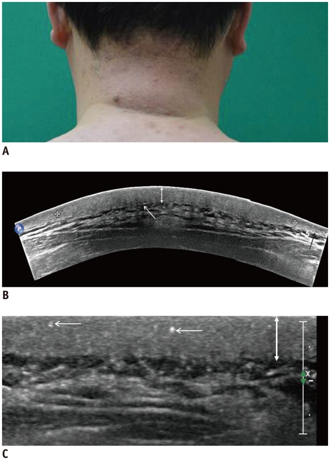
Fig. 1 44-year-old man suffering from skin thickening and palpable mass on posterior neck for 1 year.A. Photograph showing swelling and erythematous change of skin. B. Extended-field-of-view scanning image showing thickening of dermis (double-headed arrow), compared to adjacent control side (black arrow). Partial posterior acoustic shadowing (white arrow) is noted. There is no focal mass or subcutaneous fat edema. C. Multiple echogenic spots (arrows) are observed in thickened dermis (doubleheaded arrow) on ultrasonographic image.
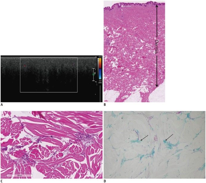
Fig. 2 46-year-old man suffering from diffuse nuchal skin thickening for 5 months.He was diagnosed with diabetes 5 years ago. A. Ultrasonographic image showing thickened dermis and marked posterior acoustic shadowing in lower dermis area. There is no vascularity on color Doppler imaging. B. Hematoxylin-eosin stain image with original magnification (× 10) showing thin and unaffected epidermis (arrow) and thickened dermis (double-headed arrow). C. Higher magnification (Hematoxylin eosin stain, × 400) image of lower portion of dermis. Collagen fibers appear swollen and separated by wide spaces. D. Alcian blue stain (× 400) demonstrating multifocal mucin depositions (arrows) between swollen collagen fibers.
Reference
-
1. Foti R, Leonardi R, Rondinone R, Di Gangi M, Leonetti C, Canova M, et al. Scleredema-like disorders. Autoimmun Rev. 2008; 7:331–339. PMID: 18295739.2. Fabri M, Hunzelmann N. [Differential diagnosis of scleredema and pseudoscleredema]. J Dtsch Dermatol Ges. 2007; 5:977–984. PMID: 17976138.3. Meguerditchian C, Jacquet P, Béliard S, Benderitter T, Valéro R, Carsuzza F, et al. Scleredema adultorum of Buschke: an under recognized skin complication of diabetes. Diabetes Metab. 2006; 32(5 Pt 1):481–484. PMID: 17110904.
Article4. Rho YW, Suhr KB, Lee JH, Park JK. A clinical observation of scleredema adultorum and its relationship to diabetes. J Dermatol. 1998; 25:103–107. PMID: 9563277.
Article5. Ioannidou DI, Krasagakis K, Stefanidou MP, Karampekios S, Panayiotidis J, Tosca AD. Scleredema adultorum of Buschke presenting as periorbital edema: a diagnostic challenge. J Am Acad Dermatol. 2005; 52(2 Suppl 1):41–44. PMID: 15692512.
Article6. Venencie PY, Powell FC, Su WP, Perry HO. Scleredema: a review of thirty-three cases. J Am Acad Dermatol. 1984; 11:128–134. PMID: 6736348.
Article7. Beers WH, Ince A, Moore TL. Scleredema adultorum of Buschke: a case report and review of the literature. Semin Arthritis Rheum. 2006; 35:355–359. PMID: 16765712.
Article8. Toyota T, Umezu M, Oikawa N, Sanoyama R, Suzuki S, Suzuki H, et al. Diabetic scleredema. Tohoku J Exp Med. 1983; 141:457–461. PMID: 6670101.
Article9. Krakowski A, Covo J, Berlin C. Diabetic scleredema. Dermatologica. 1973; 146:193–198. PMID: 4717483.
Article10. Cole GW, Handler SJ, Burnett K. The ultrasonic evaluation of skin thickness in scleredema. J Clin Ultrasound. 1981; 9:501–503. PMID: 6796610.
Article11. Haustein UF. Scleredema and pseudo-scleredema: uncommon presentations. Clin Dermatol. 2005; 23:480–490. PMID: 16179182.13. Gupta RA, Fiorentino D. Localized scleredema and systemic sclerosis: is there a connection? Best Pract Res Clin Rheumatol. 2007; 21:1025–1036. PMID: 18068859.14. Kurihara Y, Kokuba H, Furue M. Case of diabetic scleredema: diagnostic value of magnetic resonance imaging. J Dermatol. 2011; 38:693–696. PMID: 21729146.
Article15. Sattar MA, Diab S, Sugathan TN, Sivanandasingham P, Fenech FF. Scleroedema diabeticorum: a minor but often unrecognized complication of diabetes mellitus. Diabet Med. 1988; 5:465–468. PMID: 2970922.
Article
