Ann Dermatol.
2015 Aug;27(4):478-480. 10.5021/ad.2015.27.4.478.
Scleredema of Buschke Following Streptococcal Infection
- Affiliations
-
- 1Department of Dermatology, Ajou University School of Medicine, Suwon, Korea. maychan@ajou.ac.kr
- KMID: 2171518
- DOI: http://doi.org/10.5021/ad.2015.27.4.478
Abstract
- No abstract available.
Figure
-
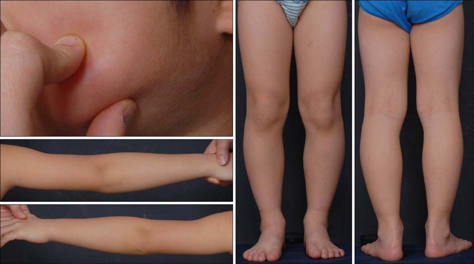
Fig. 1 Whole-body and non-pitting induration of the skin was noted including face. On physical examination, it was hard to pinch his check.
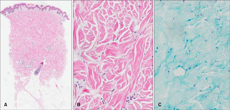
Fig. 2 (A) Skin biopsy specimen revealed a non-tapered appearance on a low-power field (H&E, ×40). (B) Fibroblasts were normal in number, and the spaces between collagen bundles were widened (H&E, ×400). (C) Mucin deposits were detected between collagen bundles in the middermis (alcian blue staining, ×400).
Reference
-
1. Weenig RH, Pittelkow MR. Scleredema and scleromyxedema. In : Goldsmith LA, Katz SI, Gilchrest BA, Paller AS, Leffell DJ, Wolff K, editors. Fitzpatrick's dermatology in general medicine. 8th ed. New York: McGraw-Hill;2012. p. 1957–1959.2. Buschke A. Ueber skleroedema. Berl Klin Wchnschr. 1902; 39:955.3. Beers WH, Ince A, Moore TL. Scleredema adultorum of Buschke: a case report and review of the literature. Semin Arthritis Rheum. 2006; 35:355–359.
Article4. Fernandez-Flores A, Gatica-Torres M, Ruelas-Villavicencio AL, Saeb-Lima M. Morphological clues in the diagnosis of sclerodermiform dermatitis. Am J Dermatopathol. 2014; 36:449–464.
Article
