A Cases of Tuberculous Pericarditis Associated with Pseudoaneurysm of the Left Ventricle
- Affiliations
-
- 1Department of Internal Medicine, Wonkwong University School of Medicine, Iri, Korea.
- KMID: 2410466
- DOI: http://doi.org/10.4250/jkse.1994.2.1.104
Abstract
- The incidence of left ventricular pseudoaneurysm is not known, but it appears to be quite rare. We experienced a case of apical pseudoaneurysm of left ventricle in a 73-year-old female who presented with progressive orthopnea. On the 2nd hospital day, cardiac tamponade developed. A small pseudoaneurysm of left ventricle with narrow neck associated with massive pericardial effusion was demonstrated by transthoracic echocardiography. Emergency coronary angiogram showed normal. Emergency operation was performed on the suspicion of rupture of the pseudoaneurysm. Microscopic examination of the wall of the aneurysm revealed fibrous tissue adhered to the granulomatous inflammatory pericardium.
MeSH Terms
Figure
-

Fig. 1. Chest PA roentgenogram shows marked cardiomegaly with well defined silhouette and blunting of both costophrenic angle with thickening of right major fissure.
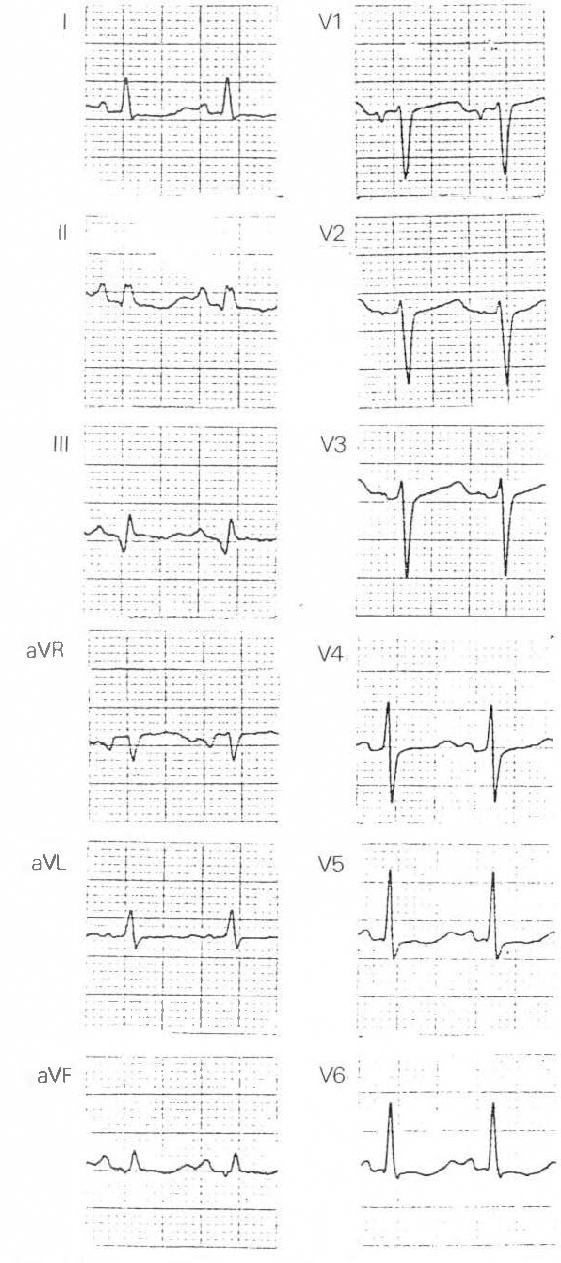
Fig. 2. Standard electrocardiogram shows sinus tachycardia with Q wave in lead II, III, aVF and diffuse non-specific ST-T change.

Fig. 3. Subcostal(A) and apical four-chamber view(B) of two-dimensional and Doppler echocardiogram show massive pericardial effusion and apical left ventricular pseudoaneurysm with narrow neck, which communicate with left ventricular cavity(P.E: pericardial effusion, RV: right ventricle, LV: left ventricle).
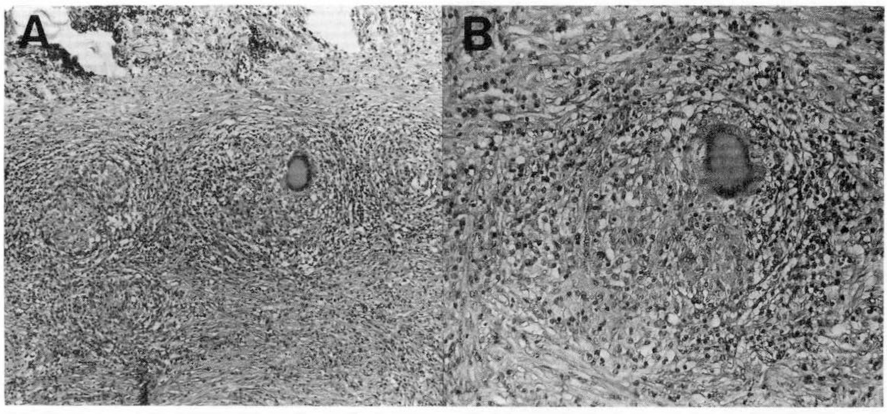
Fig. 4. Microscopic examination of the pericardium revealed granuloma with Langerhans'cell and infiltraion of inflammatory cells(H & E)(A: 100, B: 200).
Reference
-
References
1). Sabiston DC. Surgery of the chest. 5th ed.p. 1766. Philadelphia: W.B. Saunders Com;1990.2). Abrams DL. Ventricular aneurysm. Circulation. 27:164. 1963.
Article3). Stewart S. False aneurysm and pseudo-false aneurysm of the left ventricle: Etiology, pathology, diagnosis, and operative management. Ann Thorac Surg. 31:259. 1981.
Article4). Van Tassel A. Rupture of the heart complicating myocardial infarction: Analysis of 40 cases including nine examples of left ventricular false aneurysm. Chest. 61:104. 1972.5). Human DG. Tuberculous aneurysm of the left ventricle: A case report. South African Med J. 64:26. 1983.6). Salsena FB. Infective aneurysm of the left ventricle: Angio-graphic and echocardiographic features. Am Heart J. 96:385. 1978.7). Gray M, Thaker NS. Congenital calcified, apical aneurysm of the left ventricle in an adult. Eur Heart J. 8:72. 1987.
Article8). Chesler E. Subventricular and apical left ventricular aneurysms in the Bantu as a source of systemic unborn. Circulation. 35:1156. 1967.9). Arora RR, Issenberg HJ. Congenital aneurysm of the ventricle: Its recognition and significance. JAMA. 259:3306. 1988.10). Spindola-Franco H. Aneurysm of the heart: A correlative study of 102 proved cases. Medicine. 33:43. 1954.11). Spindola-Franco H. Pseudoaneurysm of the left ventricle. Radiology. 127:23. 1978.
Article12). Escarous A. CT findings of a posterior false aneurysm of the left ventricleAm J Rad. 152:1339. 1989.13). Roelandt J, Brand M. Echocardiographic diagnosis of pseudo-aneurysm of the left ventricle. Circulation. 52:466. 1975.
Article14). Glover MU, Hagan AD. Pseudoaneurysm of the left ventricule diagnosed by two-dimensional echocardiography: Case report. Military Med. 146:696. 1981.15). Grondin P, Kretz JG. Natural history of saccular aneurysms of the left ventricle. J Thor Card Surg. 77:57. 1979.
Article16). Vlodaver Z. True and false left ventricular aneurysm. Circulation. 51:567. 1975.
- Full Text Links
-
- Actions
-
Cited
- CITED
-
- Close
- Share
-
- Similar articles
-
- Tuberculous Pericarditis Mimicking a Malignant Pericardial Tumor: A Case Report
- Management of Concurrent Left Ventricular Pseudoaneurysm and Mitral Annular Calcification: A Case Report
- Malignant Pericardial Mesothelioma Misdiagnosed as Constrictive Pericarditis
- The Clinical Course of Tuberculous Pericarditis in Immunocompetent Hosts Based on Serial Echocardiography
- A Case of Tuberculous Pericarditis Diagnosed by Increased ADA Activity in Pericardial Fluid
