Echocardiographic Findings in Cardiac Tumors
- Affiliations
-
- 1Department of Internal Medicine, Dong-A University Hospital, Pusan, Korea.
- KMID: 2410437
- DOI: http://doi.org/10.4250/jkse.1995.3.1.85
Abstract
- Primary tumors of the heart are uncommon and their incidence in autopsy series ranges from 0.0017 to 0.28 per cent. About 75 per cent of all cardiac tumors are histologically benign and the remainder are malignant. The majority of benign cardiac tumors are myxoma, comprising 30 to 50 per cent of the total cases in most pathological series and the majority of malignant cardiac tumors are sarcoma. But tumors metastatic to the heart are far more common than primary cardiac tumors and actually appear to be increasing in incidence because of prolonged survival of cancer patients. We experienced six cases of cardiac tumors which were diagnosed with echocardiography : 2 hepatocellular carcinoma, 1 malignant melanoma, 1 malignant fibros histiocytoma, 1 lipoma, and 1 myxoma. The mean age of the 3 men and 3 women was 50(range 28 to 71). Two patients with right atrial metastasis from hepatocellular carcinoma and one patient with metastasis from malignant melanoma expired during conservative management. Two benign cardiac tumors(1 lipoma and 1 myxoma) were successfully excised and later follow-up echocardiography showed no signs of tumor recurrence. One patient with biatrial recurrence of malignant histiocytoma was treated medically and expired 4 months later.
Keyword
MeSH Terms
Figure
-
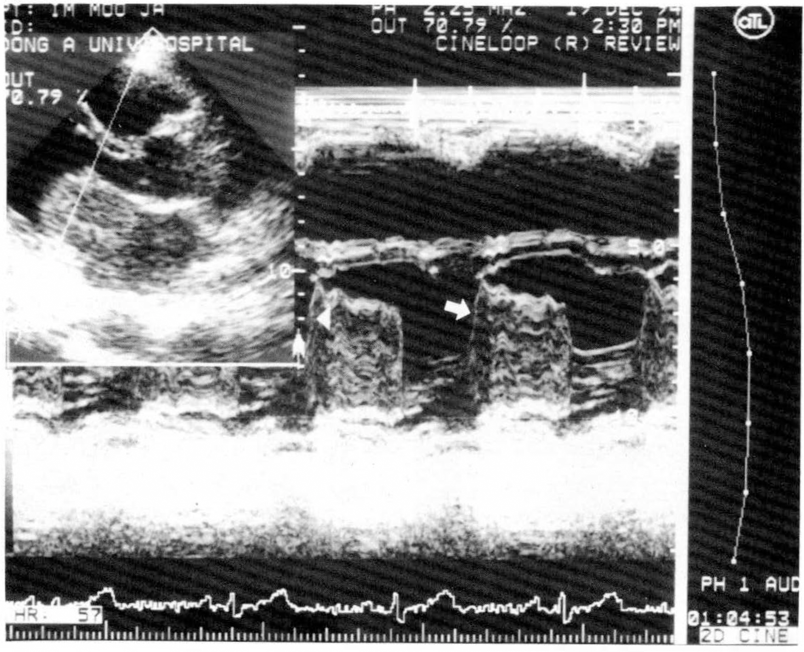
Fig. 1. Parasternal long-axis view of transthoracic echocardiogram showing 4.8 × 3.4cm sized left atrial mass attached to interatrial septum(left upper). M-mode shows tumor shadows with time gap(arrow).

Fig. 2. Echocardiographic finding of lipoma. Left: Large right atrial mass attached to atrial septum(Upper) and prolapsed into right ventricle during diastole(Lower). Right: Transesophageal echocardiogram shows 6.2 × 5.5cm sized mass with broad neck and hypertrophied interatrial septum.
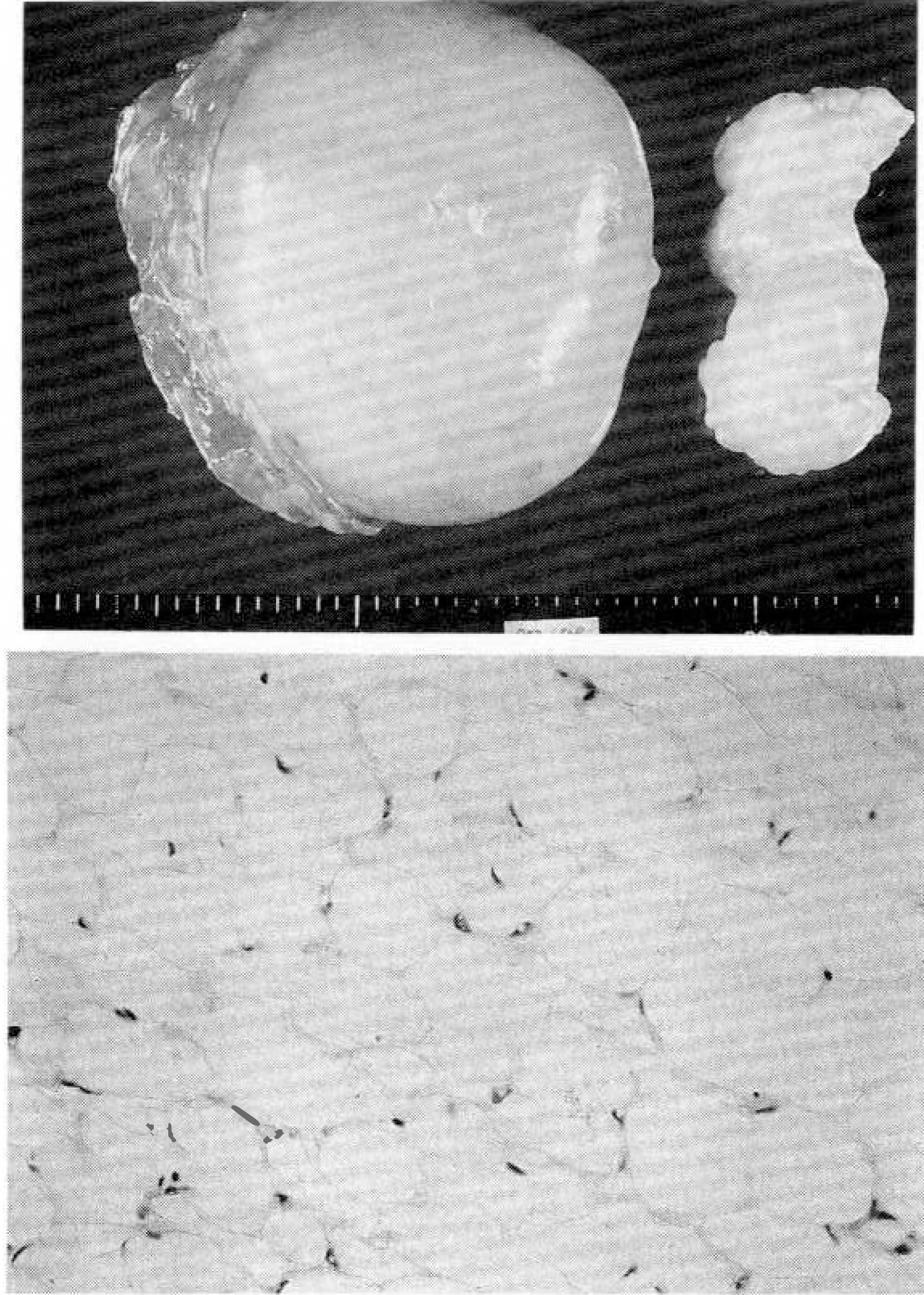
Fig. 3. Upper left: Extracardiac tumor compressing heart shows well encapsulated yellowish adipose mass with smooth and glistening surface, measuring 13.5×11 ×6cm in dimension. Upper right: Mass resected from right atrium shows well encapsulated nodular adipose mass with gray-whitish fibrous band, measuring 7×4.5×3 cm in dimension. Lower: Microscopic finding reveals lipoma that are composed of typical mature fat cells encapsulated by thin fibrous tissue.

Fig. 4. Transesophageal echocardioraphic finding shows multiple round or ovoid masses in both atria, especially filling left atrial cavity with masses and mass impinging posterior mitral valve.
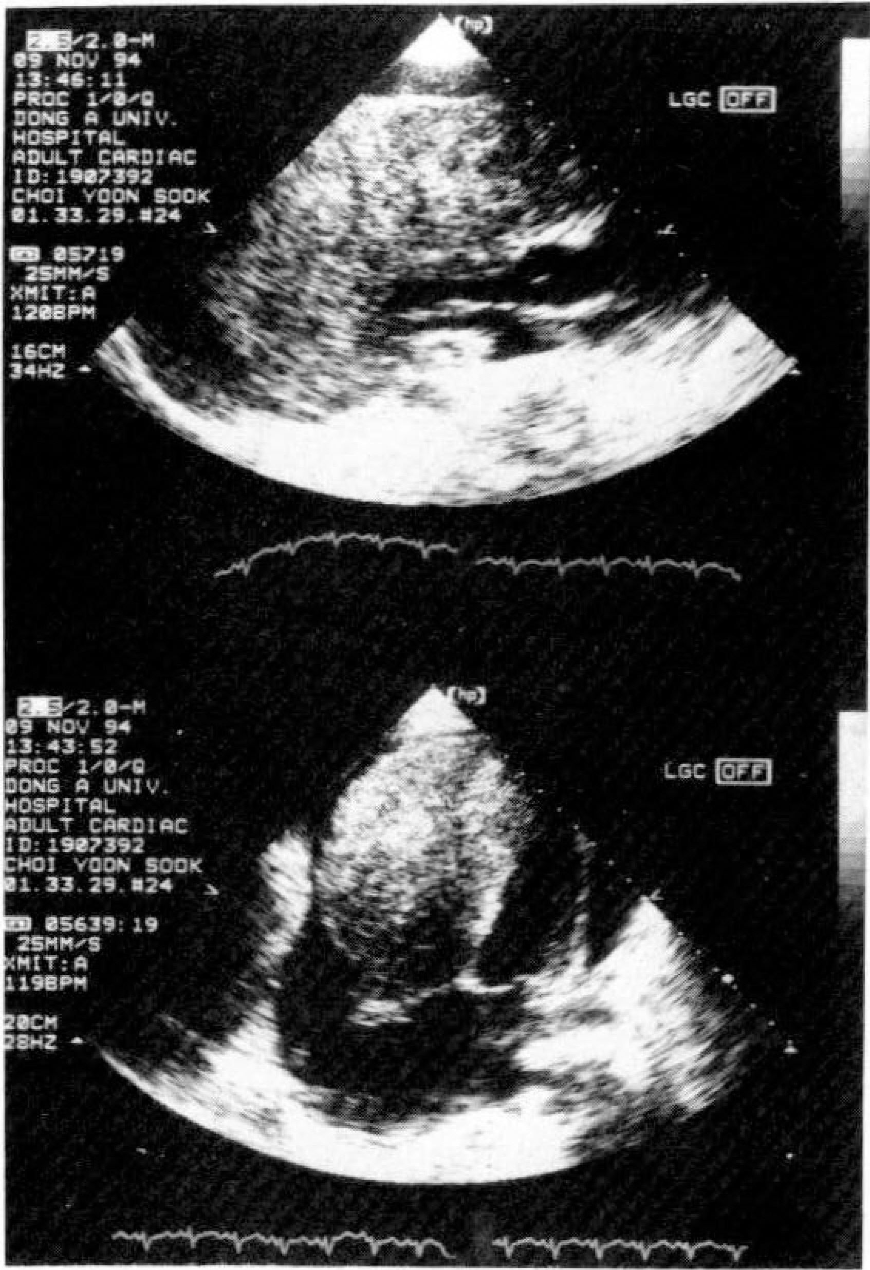
Fig. 5. Upper: Parasternal long-axis view shows mass infiltration in the nearly whole right ventricle with obstructing flow. Lower: Apical four-chamber view shows huge infiltrating mass shadow in right ventricle which invades to the interventricular septum and left ventricular wall, and massive pericardial effusion.

Fig. 6. Left: Transthoracic view shows large right atrial mass. Right: Transesophageal echocardiogram shows large mass with small daughter mass on right atrium, possibly due to cardiac metastasis.
Reference
-
References
1). Richadson JV, Brandt B III, Doty DB, Eorehaff JL. Surgical treatment of atrial myxoma: Early and late results of 11 operations and review of the literatures, Ann Thorac Surg. 28:364. 1979.2). Colucci WS, Braunwald E. Primary tumors of the heart in heart diseases. Braunwald E, editor. 4th Ed.p. 1451. Philadelphia: WB Saunders Co.;1992.3). Lammers RJ, Bloor CM. Pathology of cardiac tumors. Kapoor AS, editor. (ed.):. Cancer of the Heart. New York: Springa-Verlag;p. 62. 1986.
Article4). Bulkley BH, and Hutchins GM. Atrial myxomas: A fifty year review. Am Heart J. 97:639. 1979.
Article5). Israeli A, Rein AJ, Krivisky M, Libson E, Uziely B, and Michaeli J. Right ventricular outflow tract obstruction due to extracardiac tumors: A report of three cases diagnosed and followed up by echocardiographic studies. Arch Intern Med. 149:2105. 1989.
Article6). Kapoor AS. Clinical manifestations of neoplasia of the heart. Kapoor AS, editor. Cancer of the Heart. New York: Springa-Verlag;p. 21–25. 1986.
Article7). Schoen FJ, Berger BM, and Guerina NG. Cardiac effects of noncardiac neoplasms. Cardiol Clin. 2:657. 1984.
Article8). Barnes AR, Beaver DC, Snell AM. Primary sarcoma of the heart: Report of a case with electrocardiographic and pathological studies. Ann Heart J. 9:480. 1934.9). Goldberg HP, Glena F, Dotter CT. Myxoma of the left atrium: Diagnosis made during life with operation and postmortem findings, Circulation. 6:762. 1952.10). Seward JB, Khandheria BK, Oh JK, Abel MD. Hughes RW, Edwards WD, Nichols BA, Freeman WK, and Tajik AJ. Transesophageal echocardiography: Technique, anatomic correlations, implementation, and clinical applications. Mayo Clin Proc. 63:649–80. 1988.
Article11). Smith JS, Cahalan MK, Benefiel DJ, Byrd BF, Lurz FW, Shapiro WA, Roizen MF, Bouchard A, and Schiller NB. Intraoperative detection of myocardial ischemia in high-risk patients: Electrocardiography versus two-Dimensional transesophageal echocardiography. Circulation. 72:1015–21. 1985.
Article12). Rosenthal DS, Braunwald E. Hematological-Oncological disorders and heart diseases. Braunwald E, editor. 4th Ed.p. 1742. Philadelphia: WB Saunders Co.;1992.13). Roberts WC, Glancy DL, and DeVita VT. Heart in malignant lymphoma. A study of 196 autopsy cases. Am J Cardiol. 22:85. 1968.14). 김병주 • 왕영펼 · 곽문섭 · 김세화 • 이홍균. 심상 종양 6례 보고, 대한 흉부외과학회지.15). Kralstein J, and Frishman W. Malignant pericardial diseases: Diagnosis and tratment. Am Heart J. 113:785. 1987.16). Lloyd EA, and Curcio CA. Lymphoma of the heart as an unusual cause of pericardial effusion. S Afr Med J. 58:937. 1980.17). Haedersdal C, Hasselbach H, Devantier A, and Saunamak K. Pericardial hematopoiesis with tamponade in myelofibrosus.18). Ernesto ES, Gerald IC, Richard DW, Malcolm BD. Cardiac tumors: Diagnosis and management. Current Problems in Cardiology. 17:77–137. 1992.
Article19). Kim RH, Mautner L, Henning J. An unusual case of thyroid carcinoma with direct extension to greater veins, right heart and pulmonary arteries. Can Med Assoc J. 94:238. 1966.20). Shah AA, Churg A, Sborbarao JA, Sheppard JM, Lambert J. Malignant fibrous histiocytoma of heart presenting as an atrial myxoma. Cancer. 42:2466–2471. 1978.21). 김댁진 • 김광택 • 김영묵 • 원남회 • 안태훈 • 노영 무. 화심방내에 발생한 악성 섬유성 조직구종. 대한 흉부외과학회지. 24:357–360. 1991.22). 박종원 • 박상섭 • 류지윤 • 박현호 • 우종수. 심장 내 악성 섬유성 조직구종. 대한 흉부외과학회지. 22:297–304. 1989.23). McAllister HA, Fenoglio JJ. Tumor of the cardiovascular system. Atlas of tumor pathology, series 2. Armed Forces Institute of Pathology;1978.24). Kearney MM, Soule EH, Ivins JL. Malignant fibrous histiocytosis: A review of 167 cases. Cancer. 45:167–178. 1980.25). Fukumitsu T, Tsunekawa A, Watanabe M, Iwase M, Yakeuchi E, and Abe T. Primary malignant fibrous histiocytoma of the left atrium with acute mitral regurgitation. Am Heart J. 115:691–693. 1988.
Article26). Terashima K, Aoyama K, Nihei K, Imai Y, Takahashi K, and Daidoji S. Malignant fibrous histiocytoma of the heart. Cancer. 52:1919–26. 1983.
Article27). McCarthy PM, Piehler JM, Schaff HV, Pluth JR, Orszulak TA, Vidaillet HJ, and Carney JA. The significance of multiple, recurrent, and “complex” cardiac myxomas. Thorac Cardiovasc Surg. 91:389–396. 1986.
Article28). Sitanen P, Touteri L, Norio R, Ahrenberg P, and Halonen PI. Atrial myxoma in a family. AMJ Cardiol. 38:252. 1979.29). Goodwin JF. Diagnosis of left atrial myxoma. Lancet. 1:464. 1963.
Article30). Zitnik RS, Giuliani ER. Clinical recognition of Atrial Myxoma. Am Heart J. 80:689. 1970.
Article31). 영욱 • 이영균 · 심장 정액종. 대한 흉부외과학회지. 15:98. 1982.32). Silverman NA. Primary cardiac tumors. Ann Surg. 191:127. 1979.33). Neil Dashkoff, Ronald BV, Novin CN, Raymond G, Murray NA, Subramanian S. Bilateral atrial myxoma. Echocardiographic Consideration. Am J Med. 65:361. 1987.34). Greenwood WF. Profile of atrial myxoma. Am J Cardiol. 21:367. 1968.
Article35). Fine G. Neoplasms of the pericardium and heart. Gould SE, editor. Pathology of the heart and blood vessels. Springfield, III: Charles C Thomas;p. 851. 1968.36). Prior JT. Lipomatous hypertrophy of cardiac interatrial septum. Arch pathol. 78:11. 1964.37). Fyke FE, Tajik AJ, Edwards WD, Seward JB. Diagnosis of lipomatous hypertrophy of the atrial septum by two-dimensional echocardiography. J Am Coll Cardiol. 1:1352–57. 1983.
Article38). Keller H, Stegaru B, Buss J, Genth K, Heene D. Pulmonary tumor embolism and right atrial myxoma detected by two-dimensional echocardiography. Am Heart J. 110:881. 1985.
Article39). Currie PJ. Transesophageal echocardiography. New window to the heart. Circulation. 80:215. 1989.
Article40). Mugge A, Daniel WG, Haverich A, and Lichtlen PR. Diagnosis of noninfective cardiac mass lesions by two-dimensional echocardiography: Comparison of the transthoracic and transesophageal approaches. Circulation. 83:70–78. 1991.
Article41). Pavlides GS, Hauser AM, Stewart JR, O'Neill WW, and Timmis GC. Contribution of transesophageal echocardiography to patient diagnosis and tratment: A prospective analysis. Am Heart J. 120:910–914. 1990.42). HSU TL, Hsiung MC, Lin SL, Chen CC, Wang SP, Chang MS, and Chiang BN. The value of transesophageal echocardiography in the diagnosis of cardiac metastasis. Echocardiography. 9:1–7. 1992.
Article43). Gowin J, Axed L, Adams JR, Schiller NB, Simpson PC, Gertz EW. Computed tomography: A new method for diagnosing tumor of the heart, Circulation. 63:448. 1980.
- Full Text Links
-
- Actions
-
Cited
- CITED
-
- Close
- Share
-
- Similar articles
-
- Echocardiographic Findings in Tetralogy of Fallot
- Preoperative cardiac evaluation with transthoracic echocardiography before non-cardiac surgery
- Common Atrioventricular Canal Diagnosed by Contrast Echocardiography Report of Two Cases
- Twenty Years of Clinical Experience with Cardiac Myxomas: Diagnosis, Treatment, and Follow Up
- Echocardiographic Findings of Heart Disease in Children
