J Korean Soc Echocardiogr.
1995 Jul;3(1):32-38. 10.4250/jkse.1995.3.1.32.
Assessment of Mitral Valve Area in Patients with Mitral Stenosis Using Color Doppler Proximal Isovelocity Surface Area Method
- Affiliations
-
- 1Division of Cardiology, Department of Internal Medicine, Kyung Hee University, School of Medicinen, Seoul, Korea.
- KMID: 2410430
- DOI: http://doi.org/10.4250/jkse.1995.3.1.32
Abstract
- BACKGROUND
It has been proposed recently that measuring the flow convergence region proximal to an orifice by Doppler flow mapping can provide a means of calculating regurgitant flow rate. And this method also can be used to derive cardiac output or flow rate proximal to stenotic orifices and therefore to calculate their areas by the continuity equation(area=flow rate/velocity). Applying this method in mitral stenosis would provide a unique way of validating the underlying concept because the predicted areas could be compared with those measured directly by planimetry and pressur half-time method. METHOD: We studied 50 patients with mitral stenosis using imaging and Doppler echocardiography. Doppler color flow recordings of mitral inflow were obtained from the apex, and the radius of the proximal flow convergence region was measured at its peak diastolic valve from the orifice to the first color alias along the axis of flow. Flow rate was calculated assuming uniform radial flow convergence toward the orifice, modified by a factor that accounted for the inflow funnel angle formed by the mitral leaflets. Mitral valve area was then calculated as peak flow rate divided by peak velocity by continuous-wave Doppler.
RESULTS
The calculated areas agreed well with those from comparative techniques. 1) Cross-sectional area by planimetry(y=0.38×+0.81, r=0.51, p < 0.001). 2) Area derived from the Doppler pressure half-time(y=0.35+0.55, r=0.44, p=0.002). 3) Agreement with planimetry was similar for 26 patients with mitral regurgitation and 24 without it, as well as for 34 in atrial fibrillation.
CONCLUSION
These results suggested that the proximal flow convergence concept in the clinical setting demonstrate calculating method of mietral valve area in patients with mitral stenosis.
Keyword
MeSH Terms
Figure
-

Fig. 1. Apical four chamber view in a patient illustrating the proximal isovelocity surface area in diastole. A: As flow within the left atrium moves toward the transducer, it initally appears as red at low velocities and then changes to blue as it accelerates above the selected aliasing velocity. B: Schematic actual measurements r: radius of proximal surface area,: angle of the inlet funnel
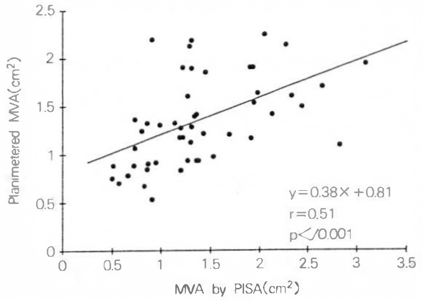
Fig. 2. Linear regression plot of mitral valve area(MVA) calculated from PISA method compared with planimetry method.
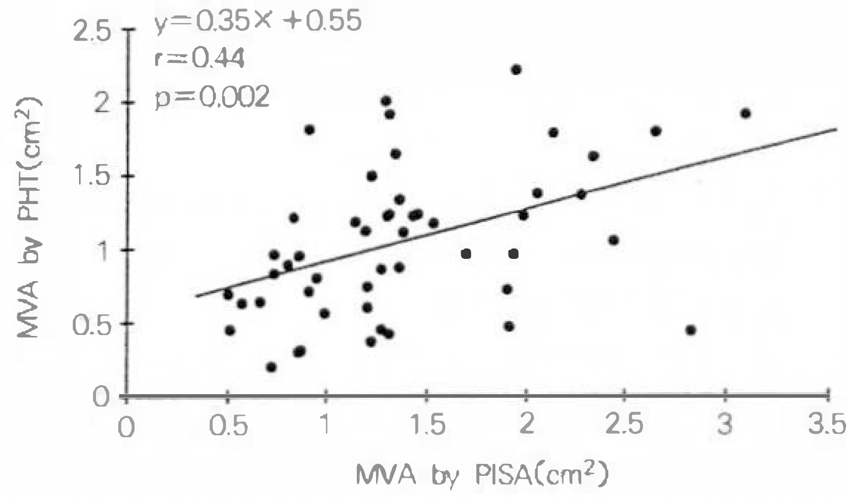
Fig. 3. Linear regression plot of mitral valve area calculated from PISA method compared with pressure half-time(PHT) method.
Reference
-
References
1). Braunwald E. Valvular heart disease. Braunwald E, editor. Heart disease: A Text book of cardiovascular Medicine. 4th ed.Philadelphia WB Saunders;1992.2). Gorlin R, Gorlin SG. Hydraulic formula for calculation of the area of stenosis mitral valve, other cardiac valves and central circulator shunts. Am heart J. 41:1–29. 1951.3). Cohn MV, Gorlin R. Modified orifice equation for the calculation of mitral valve area. Am heart J. 85:839–40. 1972.4). Nichol PM, Gilbert BW, Kisslo JA. Two-dimensional echocardiographic assessment of mitral stenosis. Circulation. 55:120–128. 1977.
Article5). Wann LS, Weymann AE, Feigenbaum H, Dill JC, Johnston KW, Eggleton RC. Determination of mitral valve area by cross-sectional echocardiography. Ann Intern Med. 88:337–341. 1978.
Article6). Martin RP, Rakowski H, Kleimal JH, Beaver W, London E, Popp RL. Reliability and reproducibility of two-dimensional echocardiographic measurement of stenotic mitral valve area. Am J Cardiol. 43:560–568. 1979.7). Smith MD, Handshoe R, Handshole S, Kwan OL, DeMaria AN. Comparative accuracy of two-dimensional echocardiography and Doppler pressure half-time method in assessing severity of mitral stenosis in patients with and without prior commissurotomy. Circulation. 73:100–107. 1986.8). Hatle L, Brubakk A, Trornsdal A, Angelsen B. Noninvasive assessment of pressure drop in mitral stenosis by Doppler ultrasound. Breart J. 40:131–40. 1978.
Article9). Halte L, Angelsen B, Trornsdal A. Noninvasive assessment of atroventricu: ar pressure half-time by Doppler ultrasound. Circulation. 60:1096–104. 1979.10). Nakatani S, Masuyama T, Kodama K, Kitabatake A, Fujii K, Kamada T. Value and limitation of Doppler echocardiography in the quantification of stenotic mitral valve area: Comparison of thee pressure-half time and the continuity equation methods. Circulation. 77:78–85. 1988.11). Flachskampf FA, Weymann AE, Gillan L, Chun-Ming L, Abascal VM, Thomas JD. Aortic regurgitation shortens Doppler pressure half-time in mitral stenosis: Clinical evidence, in vitro simulation and theoretic analysis. J Am Coll Cardiol. 16:396–404. 1990.
Article12). Karp K, Teien D, Bjerle P, Eriksson P. Reassessment of valve area determinations in mitral stenosis by the pressure half-time method: Impact of left ventricular stiffness and peak diastlic pressure difference. J Am Coll Cardiol. 13:594–9. 1989.13). Yoshida K, Yoshikava J, Yamaura Y, Hozumi T, Shakudo M, Akasaka T, Kato H. Value of acceleration flows and regurgitant jet direction by color Doppler flow mapping in the evaluation of mitral valve prolapse. Circulation. 81:879–885. 1990.
Article14). Yoshida K, Yoshikawa J, Yamamura Y, Hozumi T, Akasaka T, Iutaya T. Assessment of mitral regurgitation by biplane transesophageal color Doppler flow mapping Circulation. 82:1121–26. 1990.15). Bargiggia GS, Bertucci C, Raisaro A, Setta S, Angoli L, Recusani F, Gallati M, Tronconi L. Quantitative assessment of mitral regurgitation by color Doppler analysis of flow convergence region: Usefulness of continuity equation. Proceedings of the Sixth International Congress on Cardiac Doppler. 140. 1988.16). Bargiggia GS, Raisaro A, Bertucci C, Bramucci E, Recusani F, Montemartini C. Color Doppler analysis of proximal flow convergence region in patient with mitral regurgitation. Cardiovascular Imaging. 2:137–141. 1990.17). Gardin J. Doppler color flow “proximal isovelocity surface area(PISA)”: An alternative method for estimating volume flow across rarrowed orifice regurgitant valves and intracardiac shunt lesion. Echocardiography. 9:39–42. 1992.18). Giesler M, Scaugch M. Color Doppler determination of regurgitant flow: From proximal isovelocity surface areas to proximal velocity profiles. Echocardiography. 9:51–62. 1992.19). Utsunomiya T, Ogawa T, Patel D, et al. Doppler color flow mapping of the “proximal isovelocity surface area”. A new method for measuring volume flow across an orifice. J Am Soc Echocardiogr. 4:338–348. 1991.20). Rouse H. Fluid Mechanics for Hydraulic Engineers. New York: Dover Publications Inc.;p. 83–95. 1961.21). Utsunomiya T, Doshi R, Patel D, Mehta K, Nguyen D, Henry WL, Gardin JM. Calculation of volume flow rate by the proximal isovelocity surface area method: Simplified approach using color Doppler zero baseline shift. J Am Coll Cardiol. 22:277–82. 1993.
Article22). Utsunomiya T, Doshi R, Patel D, Nguren D, Mehta K, Gardin J. Regurgitant volume estimation in patient with mitral regurgitation initial studies using the color Doppler “proximal isovelocity surface area” method. Echocardiography. 9:63–70. 1992.23). Rodriguez L, Thomas JD, Monterrosos V, Weymann AE, Harrigan P, Mueller LN, Levine RA. Validation of the proximal flow convergence method: Calculation of orifice area in patinets with mitral stenosis. Circulation. 88:1157–1165. 1993.24). Thomas JD, wilkins GT, Choong CYP, et al. Inaccuracy of mitral pressure half-time immediately after percutaneous mitral valvotomy: dependence on transmural gradient and left atrial and ventricular compliance. Circulation. 78:980–93. 1988.25). Chen C, Wang Y, Guo B, Lin Y. Reliability of the Doppler pressure half-time method for assessing effects of percutaneous mitral balloon valvuloplasty. J Am Coll Cardiol. 13:1309–13. 1987.
Article26). Recusani F, Bargiggia GS, Yoganathan AP, Raisaro A, Valdes-Cruz LM, Sung H-W, Bertucci C, Gallati M, Moises VA, Simpson LA, Tronconi L, Sahn DJ. A new method for quantification of regurgitant flow rate using color Doppler flow imaging of the flow convergence region proximal to a discrete orifice: An in vitro study. Circulation. 83:594–604. 1991.
Article27). Utsunomiya T, Ogawa T, Doshi R, et al. Doppler color flow “proximal isovelocity surface area” method for estimating volume flow rate. Effects of orifice shape and machine factors. J Am Coll Cardiol. 17:1103. 1991.
Article28). Sahn DJ, Simpson IA, Murillo A, Valdes-Cruz L. Observation of acceleration proximal to restrictive orifices in congenital heart disease: Important clues for the interpretation of Doppler color flow maps. Circulation. 1988; 78(suppl II):II–649. Abstract.29). Moises VA, Maciel BC, Hornberger LK, Murillo-Olivas A, Valdes-Cruz LM, Sahn DJ, Weintraub RG. Anew method for noninvasive estimation of ventricular septal defect shunt flow by Doppler color flow mapping: Imaging of the laminar flow convergence region on the left septal surface. J Am Coll Cardiol. 18:824–832. 1991.30). 강흥선 • 조정휘 · 김권삼 • 김명식 • 송정상 · 배종 화. 숭모판 폐쇄부천중환자에서 색채 도플러 심초 음파도의 근위부 동속 표면적(PISA) 방볍올 이용한 역류혈액 량 측정, 한국심초옴파학회지. 2(1):41–52. 1994.
- Full Text Links
-
- Actions
-
Cited
- CITED
-
- Close
- Share
-
- Similar articles
-
- Usefulness of Pressure Half Time by Pulse Doppler Ultrasound in Evaluation of the Severity of Mitral Stenosis
- Quantitative Evaluation of Regurgitant Volume in the Patients with Mitral Regurgitation Using Color Doppler Proximal Isovelocity Surface Area Method
- Assessment of Mitral Stenosis by Doppler Echocardiography: Influence of Regurgitation on Doppler Pressure Half-Time
- Mitral Valve Area and Resistance in Mitral Stenosis: Comparison of Cardiac Catheterization and Doppler Echocardiography
- Doppler Echocardiographic Assessment of Diastolic Pressure Gradient and Mitral Valve Area in Mitral Valvular Disease
