J Korean Soc Echocardiogr.
1995 Dec;3(2):138-143. 10.4250/jkse.1995.3.2.138.
Blood Flow Pattern of Left and Right Coronary Arteries in Patients with Coronary Arterial Disease Measured by Intracoronary Doppler-tipped Guidewire
- Affiliations
-
- 1Department of Internal Medicine, College of Medicine, Dong-A University, Pusan, Korea.
- 2Department of Internal Medicine, Dae Dong Hospital, Pusan, Korea.
- KMID: 2410415
- DOI: http://doi.org/10.4250/jkse.1995.3.2.138
Abstract
- BACKGROUND
Measurement of coronary flow velocity in clinical caes contributes to understanding the pathophysiology of coronary circulation. To evaluate the coronary hemodynamics, we analyzed the pattern of coronary flow velocity with a new device consisting of 15Mhz piezoelectric transducer integrated into the tipped 0.018 inch or 0.014 inch flexible, steerable angioplasty guidewire. METHOD: A low profile(0.018 in. or 0.014 in.) Doppler angioplasty guidewire was used to measure the basal blood flow velocity in proximal coronary artery after intracoronary infusion of 200µg nitroglycerine, hyperemic blood flow velocity after intracoronary infusion of adenosine(12µg for LCA, 6µg for RCA). We measured several parameters such as APV (average peak velocity, cm/sec), DSVR(diastolic systolic velocity ratio), MPV(maximal peak velosity, cm/sec), PVI(peak velocity integral, cm), SPVI(systolic peak velocity integral, cm), DSIR(diastolic systolic integral ratio), ASPV(average systolic peak velocity, cm/sec) in basal and hyperemic states. This measurements were made in 17 patients undergoing coronary angiography.
RESULTS
1) APV, ADPV, MPV, in the basal state were higher in LCA than in RCA(32.1±16.6, 40.0±23.6, 57.1±29.0/15.8±9.1, 17.1±10.2, 24.5±20.9cm/sec) and DSVR was also higher in LCA(2.7±2.1/1.4±0.6). But ASPV and SPVI was not different(p>0.05). Significant increases in APV were noted in LCA(32.1±16.6→60.6±17.6cm/sec) and in RCA (15.8±9.1→42.1±15.5cm/sec) after adenosine infusion compared with basal state. DSVR measured in basal state were not statistically different from values in hyperemic state in LCA and RCA(2.7±2.1→2.3±1.7, 1.4±0.6→1.4±0.5, p>0.05).
CONCLUSION
The blood flow patterns in both coronary arteries showed different biphasic flow patterns and this finding might be due to the pressure gradient during diastolic phase of both ventricles.
MeSH Terms
Figure
-
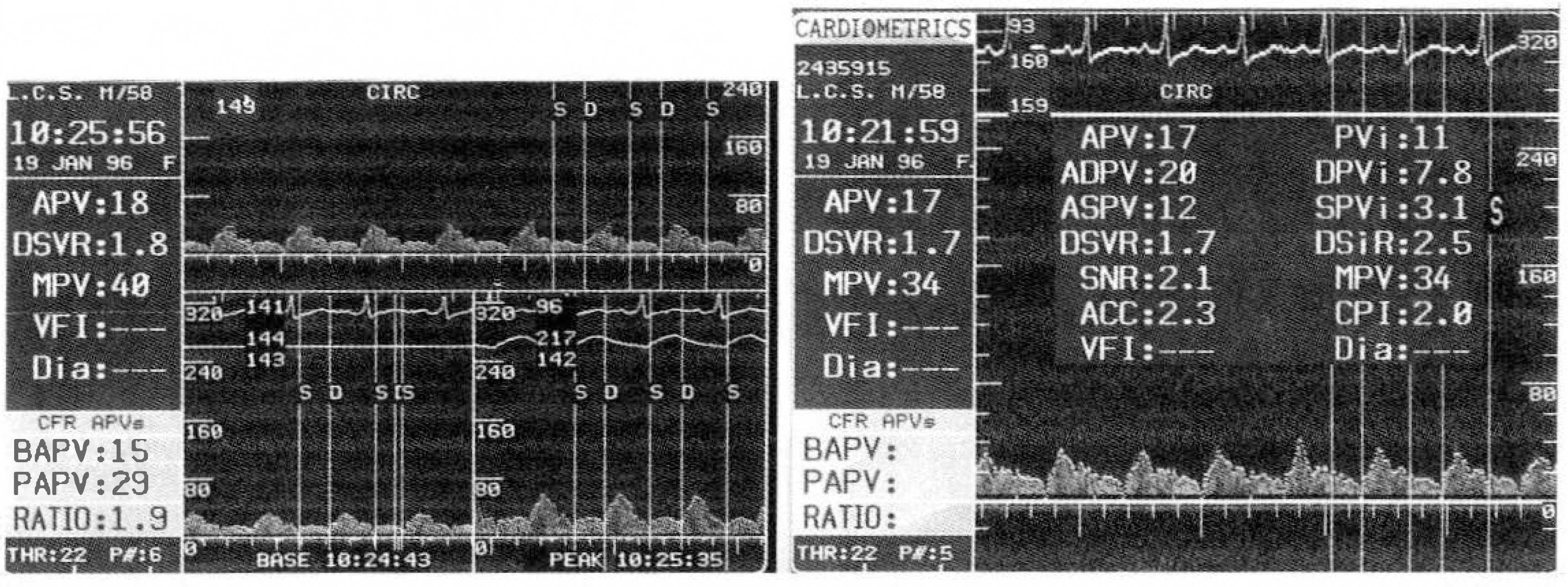
Fig. 1. Diastolic dominant flow pattern in the proximal segment of moderately stenotic left coronary artery(left) and measured several parameters(right). APV=Average peak velocity ASPV=Average systolic peak velocity PVi = Peak velocity integral SPVi=Systolic peak velocity integral ADPV=Average diastolic peak velocity DSVR=Diastolic systolic velocity ratio DPVi = Diastolic peak velocity integral DSiR= Diastolic systolic integral ratio MPV=Mean peak velocity
- Full Text Links
-
- Actions
-
Cited
- CITED
-
- Close
- Share
-
- Similar articles
-
- Coronary Flow Patterns in the Coronary Artery Narrowings
- Myocardial Contrast Echocardiography for the Assessment of Coronary Blood Flow Reserve
- Coronary Flow Velocity Pattern in Patients with Myocardial Bridging of Coronary Artery
- Phasic Coronary Artery Flow Profiles in Patients with Aortic Valve Disease
- Visualization of Coronary Arteries by Color-Coded Transesophageal Doppler Echocardiography
