Invasive Lobular Carcinoma: Detection and Multiplicity with Multimodalities
- Affiliations
-
- 1Department of Radiology, Ewha Womans University College of Medicine, Seoul, Korea. aqua0724@ewha.ac.kr
- 2Department of Radiology, National Cancer Center, Goyang, Korea.
- 3Department of Nuclear Medicine, Ewha Womans University College of Medicine, Seoul, Korea.
- KMID: 2410331
- DOI: http://doi.org/10.12771/emj.2018.41.2.27
Abstract
OBJECTIVES
We aimed to compare the diagnostic performances of digital mammography (DM), digital breast tomosynthesis (DBT), ultrasound (US), magnetic resonance imaging (MRI), breast specific gamma imaging (BSGI) and/or positron emission tomography/computed tomography (PET/CT) for the detection of invasive lobular carcinoma (ILC).
METHODS
Index ILCs and multifocal/multicentric (multiple) ILCs were analyzed using various imaging modalities. The final surgical pathology was regarded as the reference standard. The detection rate for index cancers and the diagnostic performance for multiple ILCs per breast were evaluated.
RESULTS
Seventy-eight ILCs in 76 women were enrolled. Twenty-six breasts had multiple ILCs. DM (n=72), DBT (n=15), US (n=77), MRI (n=76), BSGI (n=50), and /or PET/CT (n=74) were performed. For index cancer, the detection rate was 100% for DBT, US, and MRI. For multiple ILCs, the sensitivity was 100% for DBT and MRI (P<0.001). The diagnostic accuracy for multiple ILCs were 73.3% for DBT and 73.0% for PET/CT (P=0.460).
CONCLUSION
DBT was the most accurate imaging modality for both index and multiple ILCs. PET/CT was also valuable for multiple ILCs, whereas DM and BSGI showed relatively low diagnostic performances. DBT and PET/CT have promising roles in the diagnosis of multiple ILCs.
Keyword
MeSH Terms
Figure
-

Fig. 1 Case of a 42-year-old woman who presented for screening. (A) Digital mammography shows asymmetry (arrow) in the left breast. (B) An irregular, spiculated and hypoechoic mass was noted at the left breast. It was confirmed as an invasive lobular carcinoma by ultrasound-guided core needle biopsy. (C) Breast specific gamma imaging showed a focal faint radiotracer uptake (arrow) uptake in the left inner breast. (D) Positron emission tomography image does not show abnormal fludeoxyglucose uptake in the left breast. (E) Dynamic contrast enhanced magnetic resonance imaging show a rim enhancing mass (arrow) in left breast.
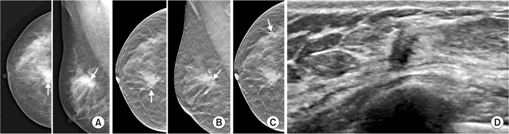
Fig. 2 A 45-year-old woman with known invasive lobular carcinoma in right breast. (A) Right craniocaudal and mediolateral oblique views of digital mammography and (B) digital breast tomosynthesis images revealing known index cancer (arrows) in right upper central breast. (C) The spiculated margin of index cancer is more clearly seen on digital breast tomosynthesis images than digital mammography. Different level image of tomosynthesis shows an additional asymmetry (arrow) in right outer breast, which is not depicted on digital mammography. (D) An irregular, spiculated and hypoechoic mass was noted at 9 o'clock of right breast on breast ultrasound. It was proven as an additional invasive lobular carcinoma by ultrasound-guided core needle biopsy.
Reference
-
1. Li CI, Anderson BO, Daling JR, Moe RE. Trends in incidence rates of invasive lobular and ductal breast carcinoma. JAMA. 2003; 289:1421–1424.
Article2. Arpino G, Bardou VJ, Clark GM, Elledge RM. Infiltrating lobular carcinoma of the breast: tumor characteristics and clinical outcome. Breast Cancer Res. 2004; 6:R149–R156.
Article3. Glass AG, Lacey JV Jr, Carreon JD, Hoover RN. Breast cancer incidence, 1980-2006: combined roles of menopausal hormone therapy, screening mammography, and estrogen receptor status. J Natl Cancer Inst. 2007; 99:1152–1161.4. Biglia N, Mariani L, Sgro L, Mininanni P, Moggio G, Sismondi P. Increased incidence of lobular breast cancer in women treated with hormone replacement therapy: implications for diagnosis, surgical and medical treatment. Endocr Relat Cancer. 2007; 14:549–567.
Article5. Korhonen T, Huhtala H, Holli K. A comparison of the biological and clinical features of invasive lobular and ductal carcinomas of the breast. Breast Cancer Res Treat. 2004; 85:23–29.
Article6. Qureshi HS, Linden MD, Divine G, Raju UB. E-cadherin status in breast cancer correlates with histologic type but does not correlate with established prognostic parameters. Am J Clin Pathol. 2006; 125:377–385.
Article7. Derksen PW, Braumuller TM, van der, Hornsveld M, Mesman E, Wesseling J, et al. Mammary-specific inactivation of E-cadherin and p53 impairs functional gland development and leads to pleomorphic invasive lobular carcinoma in mice. Dis Model Mech. 2011; 4:347–358.
Article8. Evans WP, Warren Burhenne LJ, Laurie L, O'Shaughnessy KF, Castellino RA. Invasive lobular carcinoma of the breast: mammographic characteristics and computer-aided detection. Radiology. 2002; 225:182–189.
Article9. Krecke KN, Gisvold JJ. Invasive lobular carcinoma of the breast: mammographic findings and extent of disease at diagnosis in 184 patients. AJR Am J Roentgenol. 1993; 161:957–960.
Article10. Veltman J, Boetes C, van Die L, Bult P, Blickman JG, Barentsz JO. Mammographic detection and staging of invasive lobular carcinoma. Clin Imaging. 2006; 30:94–98.
Article11. Le Gal M, Ollivier L, Asselain B, Meunier M, Laurent M, Vielh P, et al. Mammographic features of 455 invasive lobular carcinomas. Radiology. 1992; 185:705–708.
Article12. Paramagul CP, Helvie MA, Adler DD. Invasive lobular carcinoma: sonographic appearance and role of sonography in improving diagnostic sensitivity. Radiology. 1995; 195:231–234.
Article13. Selinko VL, Middleton LP, Dempsey PJ. Role of sonography in diagnosing and staging invasive lobular carcinoma. J Clin Ultrasound. 2004; 32:323–332.
Article14. Mann RM, Hoogeveen YL, Blickman JG, Boetes C. MRI compared to conventional diagnostic work-up in the detection and evaluation of invasive lobular carcinoma of the breast: a review of existing literature. Breast Cancer Res Treat. 2008; 107:1–14.
Article15. Johnson K, Sarma D, Hwang ES. Lobular breast cancer series: imaging. Breast Cancer Res. 2015; 17:94.
Article16. Friedewald SM, Rafferty EA, Rose SL, Durand MA, Plecha DM, Greenberg JS, et al. Breast cancer screening using tomosynthesis in combination with digital mammography. JAMA. 2014; 311:2499–2507.
Article17. Brem RF, Ioffe M, Rapelyea JA, Yost KG, Weigert JM, Bertrand ML, et al. Invasive lobular carcinoma: detection with mammography, sonography, MRI, and breast-specific gamma imaging. AJR Am J Roentgenol. 2009; 192:379–383.
Article18. Jung NY, Kim SH, Kim SH, Seo YY, Oh JK, Choi HS, et al. Effectiveness of breast MRI and (18)F-FDG PET/CT for the preoperative staging of invasive lobular carcinoma versus ductal carcinoma. J Breast Cancer. 2015; 18:63–72.19. Mariscotti G, Durando M, Houssami N, Zuiani C, Martincich L, Londero V, et al. Digital breast tomosynthesis as an adjunct to digital mammography for detecting and characterising invasive lobular cancers: a multi-reader study. Clin Radiol. 2016; 71:889–895.
Article20. Destounis SV, Morgan R, Arieno A. Screening for dense breasts: digital breast tomosynthesis. AJR Am J Roentgenol. 2015; 204:261–264.
Article21. Hooley RJ, Greenberg KL, Stackhouse RM, Geisel JL, Butler RS, Philpotts LE. Screening US in patients with mammographically dense breasts: initial experience with Connecticut Public Act 09-41. Radiology. 2012; 265:59–69.
Article22. Skaane P, Bandos AI, Gullien R, Eben EB, Ekseth U, Haakenaasen U, et al. Comparison of digital mammography alone and digital mammography plus tomosynthesis in a populationbased screening program. Radiology. 2013; 267:47–56.
Article23. Goldsmith SJ, Parsons W, Guiberteau MJ, Stern LH, Lanzkowsky L, Weigert J, et al. SNM practice guideline for breast scintigraphy with breast-specific gamma-cameras 1.0. J Nucl Med Technol. 2010; 38:219–224.24. Berg WA, Gutierrez L, NessAiver MS, Carter WB, Bhargavan M, Lewis RS, et al. Diagnostic accuracy of mammography, clinical examination, US, and MR imaging in preoperative assessment of breast cancer. Radiology. 2004; 233:830–849.
Article25. Hilleren DJ, Andersson IT, Lindholm K, Linnell FS. Invasive lobular carcinoma: mammographic findings in a 10-year experience. Radiology. 1991; 178:149–154.
Article26. Lee WK, Chung J, Cha ES, Lee JE, Kim JH. Digital breast tomosynthesis and breast ultrasound: additional roles in dense breasts with category 0 at conventional digital mammography. Eur J Radiol. 2016; 85:291–296.
Article27. Noroozian M, Hadjiiski L, Rahnama-Moghadam S, Klein KA, Jeffries DO, Pinsky RW, et al. Digital breast tomosynthesis is comparable to mammographic spot views for mass characterization. Radiology. 2012; 262:61–68.
Article28. Buck AK, Schirrmeister H, Mattfeldt T, Reske SN. Biological characterisation of breast cancer by means of PET. Eur J Nucl Med Mol Imaging. 2004; 31:Suppl 1. S80–S87.
Article29. Kim BS. Usefulness of breast-specific gamma imaging as an adjunct modality in breast cancer patients with dense breast: a comparative study with MRI. Ann Nucl Med. 2012; 26:131–137.
Article30. Kelley KA, Crawford JD, Thomas K, Gardiner SK, Johnson NG. A comparison of breast-specific gamma imaging of invasive lobular carcinomas and ductal carcinomas. JAMA Surg. 2015; 150:816–818.
Article
- Full Text Links
-
- Actions
-
Cited
- CITED
-
- Close
- Share
-
- Similar articles
-
- Nodular Metastatic Carcinoma from Invasive Lobular Breast Cancer
- Invasive Lobular Carcinoma of the Breast Associated with Mixed Lobular and Ductal Carcinoma In Situ: A Case Report
- Lobular carcinoma in situ in sclerosing adenosis
- Invasive Lobular Carcinoma Mimicking Fat Necrosis in Breast: A Case Report
- Signet Ring Cell Variant of Invasive Lobular Carcinoma of Male Breast
