Imaging Sci Dent.
2016 Sep;46(3):179-184. 10.5624/isd.2016.46.3.179.
The use of digital periapical radiographs to study the prevalence of alveolar domes
- Affiliations
-
- 1Department of Oral Radiology, School of Dentistry, Pontifical Catholic University of Minas Gerais, Belo Horizonte, Brazil. manzi@pucminas.br
- KMID: 2408250
- DOI: http://doi.org/10.5624/isd.2016.46.3.179
Abstract
- PURPOSE
In the present study, we coined the term 'alveolar dome' and aimed to demonstrate the prevalence of alveolar domes through digital periapical radiographs.
MATERIALS AND METHODS
This study examined 800 digital periapical radiographs in regard to the presence of alveolar domes. The periapical radiographs were acquired by a digital system using a photostimulable phosphor (PSP) plate. The χ2 test, with a significance level of 5%, was used to compare the prevalence of alveolar domes in the maxillary posterior teeth and, considering the same teeth, to verify the difference in the prevalence of dome-shaped phenomena between the roots.
RESULTS
The prevalence of alveolar domes present in the first pre-molars was statistically lower as compared to the other maxillary posterior teeth (p<0.05). No statistically significant difference was observed in the prevalence of alveolar domes between the maxillary first and second molars. Considering the maxillary first and second molars, it was observed that the palatal root presented a lower prevalence of alveolar domes when compared to the distobuccal and mesiobuccal roots (p<0.05).
CONCLUSION
The present study coined the term 'alveolar dome', referring to the anatomical projection of the root into the floor of the maxillary sinus. The maxillary first and second molars presented a greater prevalence of alveolar domes, especially in the buccal roots, followed by the third molars and second pre-molars. Although the periapical radiograph is a two-dimensional method, it can provide dentists with the auxiliary information necessary to identify alveolar domes, thus improving diagnosis, planning, and treatment.
MeSH Terms
Figure
-

Fig. 1 Maxillary sinus without pneumatization. The periapical radiograph shows the slightly curved, thin, delicate, and tenuous radiopaque line of the contour of the maxillary sinus floor (arrows).
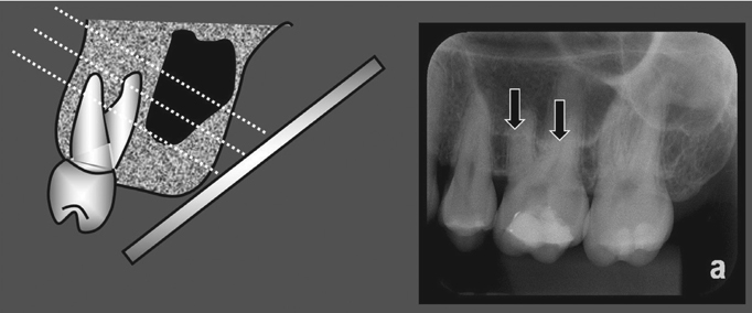
Fig. 2 Maxillary sinus with pneumatization near the root of the maxillary molar. The contour of the maxillary sinus floor is projected over the roots of the maxillary molar; however, its format remains horizontal and slightly curved (arrows).
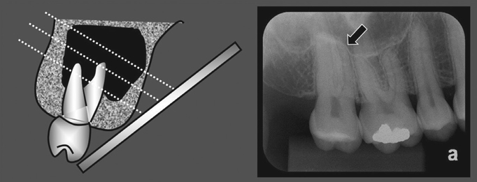
Fig. 3 Maxillary sinus with pneumatization involving the root of the maxillary molar. The radiopaque line of the contour of the maxillary sinus floor appears in the form of a bell, forming an alveolar dome (arrows).
Reference
-
1. Hauman CH, Chandler NP, Tong DC. Endodontic implications of the maxillary sinus: a review. Int Endod J. 2002; 35:127–141.
Article2. Pagin O, Centurion BS, Rubira-Bullen IR, Alvares Capelozza AL. Maxillary sinus and posterior teeth: accessing close relationship by cone-beam computed tomographic scanning in a Brazilian population. J Endod. 2013; 39:748–751.
Article3. Kilic C, Kamburoglu K, Yuksel SP, Ozen T. An assessment of the relationship between the maxillary sinus floor and the maxillary posterior teeth root tips using dental cone-beam computerized tomography. Eur J Dent. 2010; 4:462–467.
Article4. de Oliveira AG, dos Santos Silveira O, Francio LA, de Andrade Marigo Grandinetti H, Manzi FR. Anatomic variations of paranasal sinuses - clinical case report. Surg Radiol Anat. 2013; 35:535–538.5. Lana JP, Carneiro PM, Machado Vde C, de Souza PE, Manzi FR, Horta MC. Anatomic variations and lesions of the maxillary sinus detected in cone beam computed tomography for dental implants. Clin Oral Implants Res. 2012; 23:1398–1403.6. Sharan A, Madjar D. Correlation between maxillary sinus floor topography and related root position of posterior teeth using panoramic and cross-sectional computed tomography imaging. Oral Surg Oral Med Oral Pathol Oral Radiol Endod. 2006; 102:375–381.
Article7. Kretzschmar DP, Kretzschmar JL. Rhinosinusitis: review from a dental perspective. Oral Surg Oral Med Oral Pathol Oral Radiol Endod. 2003; 96:128–135.
Article8. Tan R, Spector S. Pediatric sinusitis. Curr Allergy Asthma Rep. 2007; 7:421–426.
Article9. Patel NA, Ferguson BJ. Odontogenic sinusitis: an ancient but under-appreciated cause of maxillary sinusitis. Curr Opin Otolaryngol Head Neck Surg. 2012; 20:24–28.10. Pokorny A, Tataryn R. Clinical and radiologic findings in a case series of maxillary sinusitis of dental origin. Int Forum Allergy Rhinol. 2013; 3:973–979.
Article11. Didilescu A, Rusu M, Săndulescu M, Georgescu C, Ciuluvică R. Morphometric analysis of the relationships between the maxillary first molar and maxillary sinus floor. Open J Stomatol. 2012; 2:352–357.
Article12. Eberhardt JA, Torabinejad M, Christiansen EL. A computed tomographic study of the distances between the maxillary sinus floor and the apices of the maxillary posterior teeth. Oral Surg Oral Med Oral Pathol. 1992; 73:345–346.
Article13. Kwak HH, Park HD, Yoon HR, Kang MK, Koh KS, Kim HJ. Topographic anatomy of the inferior wall of the maxillary sinus in Koreans. Int J Oral Maxillofac Surg. 2004; 33:382–388.
Article14. American Dental Association, U.S. Department of Health and Human Services. Dental radiographic examinations: recommendations for patient selection and limiting radiation exposure. Revised: 2012 [Internet]. Chicago: American Dental Association;2012. cited 2016 Mar 1. Available from: http://www.ada.org/~/media/ADA/Member Center/FIles/Dental_Radiographic_Examinations_2012.ashx.15. Low KM, Dula K, Bürgin W, von Arx T. Comparison of periapical radiography and limited cone-beam tomography in posterior maxillary teeth referred for apical surgery. J Endod. 2008; 34:557–562.
Article16. White SC, Pharoah MJ. Oral radiology; principles and interpretation. 6th ed. St. Louis: Mosby-Year Book Inc;2009.17. Shanbhag S, Karnik P, Shirke P, Shanbhag V. Association between periapical lesions and maxillary sinus mucosal thickening: a retrospective cone-beam computed tomographic study. J Endod. 2013; 39:853–857.
Article18. Lu Y, Liu Z, Zhang L, Zhou X, Zheng Q, Duan X, et al. Associations between maxillary sinus mucosal thickening and apical periodontitis using cone-beam computed tomography scanning: a retrospective study. J Endod. 2012; 38:1069–1074.
Article19. Mossa-Basha M, Blitz AM. Imaging of the paranasal sinuses. Semin Roentgenol. 2013; 48:14–34.
Article20. Lane JJ, O'Neal RB. The relationship between periodontitis and the maxillary sinus. J Periodontol. 1984; 55:477–481.
Article21. Butaric LN, McCarthy RC, Broadfield DC. A preliminary 3D computed tomography study of the human maxillary sinus and nasal cavity. Am J Phys Anthropol. 2010; 143:426–436.
Article22. Jung YH, Cho BH. Assessment of the relationship between the maxillary molars and adjacent structures using cone beam computed tomography. Imaging Sci Dent. 2012; 42:219–224.
Article23. Special Committee to Revise the Joint AAE/AAOMR Position Statement on use of CBCT in Endodontics. AAOMR Position Statement on use of CBCT in Endodontics. AAE and AAOMR joint position statement: use of cone beam computed tomography in endodontics 2015 update. Oral Surg Oral Med Oral Pathol Oral Radiol. 2015; 120:508–512.
- Full Text Links
-
- Actions
-
Cited
- CITED
-
- Close
- Share
-
- Similar articles
-
- An experimental study on the readability of digital images in the furcal bone defects
- Comparison of digital radiometric featuresbetween radicular cysts and periapical granulomas
- Image enhancement of digital periapical radiographs according to diagnostic tasks
- Prediction of osteoporosis using fractal analysis on periapical and panoramic radiographs
- Diagnostic accuracy of artificially induced vertical root fractures: a comparison of direct digital periapical images with conventional periapical images
