Imaging Sci Dent.
2014 Mar;44(1):31-35. 10.5624/isd.2014.44.1.31.
Image enhancement of digital periapical radiographs according to diagnostic tasks
- Affiliations
-
- 1Department of Oral and Maxillofacial Radiology, Dankook University College of Dentistry, Cheonan, Korea. ekkim@dankook.ac.kr
- KMID: 1974471
- DOI: http://doi.org/10.5624/isd.2014.44.1.31
Abstract
- PURPOSE
This study was performed to investigate the effect of image enhancement of periapical radiographs according to the diagnostic task.
MATERIALS AND METHODS
Eighty digital intraoral radiographs were obtained from patients and classified into four groups according to the diagnostic tasks of dental caries, periodontal diseases, periapical lesions, and endodontic files. All images were enhanced differently by using five processing techniques. Three radiologists blindly compared the subjective image quality of the original images and the processed images using a 5-point scale.
RESULTS
There were significant differences between the image quality of the processed images and that of the original images (P<0.01) in all the diagnostic task groups. Processing techniques showed significantly different efficacy according to the diagnostic task (P<0.01).
CONCLUSION
Image enhancement affects the image quality differently depending on the diagnostic task. And the use of optimal parameters is important for each diagnostic task.
Figure
-
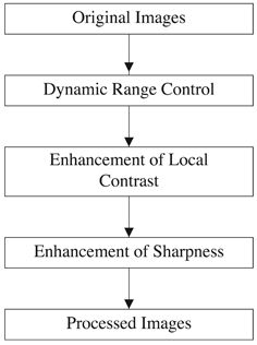
Fig. 1 Stages of image enhancement.
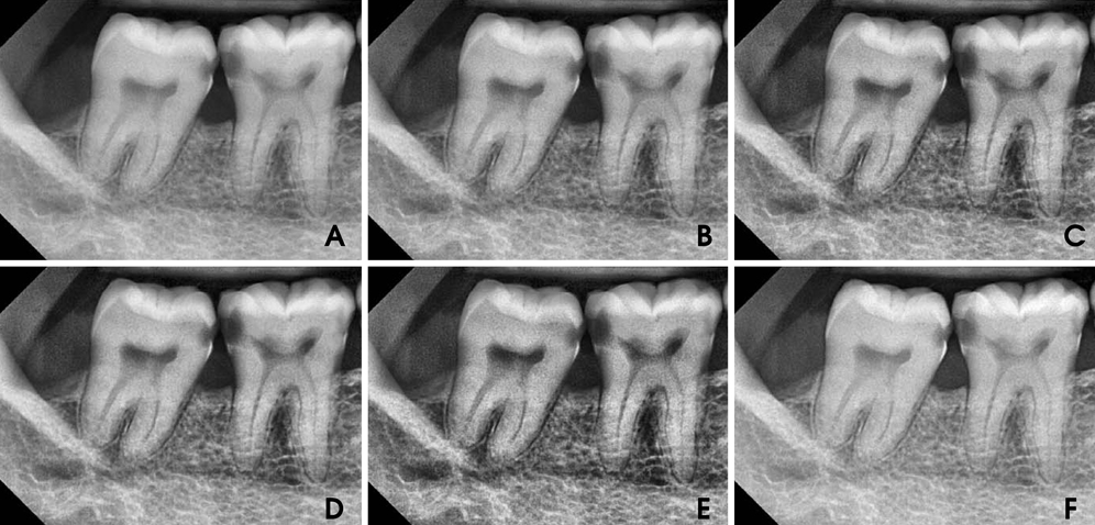
Fig. 2 Original and processed images. A. image enhanced by processing technique A. B. image enhanced by processing technique B. C. image enhanced by processing technique C. D. image enhanced by processing technique D. E. image enhanced by processing technique E. F. original image.
Reference
-
1. Körner M, Weber CH, Wirth S, Pfeifer KJ, Reiser MF, Treitl M. Advances in digital radiography: physical principles and system overview. Radiographics. 2007; 27:675–686.
Article2. Wenzel A. Current trends in radiographic caries imaging. Oral Surg Oral Med Oral Pathol Oral Radiol Endod. 1995; 80:527–539.
Article3. Farman AG, Avant SL, Scarfe WC, Farman TT, Green DB. In vivo comparison of Visualix-2 and Ektaspeed Plus in the assessment of periradicular lesion dimensions. Oral Surg Oral Med Oral Pathol Oral Radiol Endod. 1998; 85:203–209.
Article4. Sund T, Møystad A. Sliding window adaptive histogram equalization of intraoral radiographs: effect on image quality. Dentomaxillofac Radiol. 2006; 35:133–138.
Article5. Møystad A, Svanaes DB, Risnes S, Larheim TA, Gröndahl HG. Detection of approximal caries with a storage phosphor system. A comparison of enhanced digital images with dental X-ray film. Dentomaxillofac Radiol. 1996; 25:202–206.
Article6. Li G, Yoshiura K, Welander U, Shi XQ, McDavid WD. Detection of approximal caries in digital radiographs before and after correction for attenuation and visual response. An in vitro study. Dentomaxillofac Radiol. 2002; 31:113–116.
Article7. Shi XQ, Li G. Detection accuracy of approximal caries by black-and-white and color-coded digital radiographs. Oral Surg Oral Med Oral Pathol Oral Radiol Endod. 2009; 107:433–436.
Article8. Scarfe WC, Czerniejewski VJ, Farman AG, Avant SL, Molteni R. In vivo accuracy and reliability of color-coded image enhancements for the assessment of periradicular lesion dimensions. Oral Surg Oral Med Oral Pathol Oral Radiol Endod. 1999; 88:603–611.
Article9. Kullendorff B, Petersson K, Rohlin M. Direct digital radiography for the detection of periapical bone lesions: a clinical study. Endod Dent Traumatol. 1997; 13:183–189.
Article10. Dove SB, McDavid WD. A comparison of conventional intraoral radiography and computer imaging techniques for the detection of proximal surface dental caries. Dentomaxillofac Radiol. 1992; 21:127–134.
Article11. Tyndall DA, Ludlow JB, Platin E, Nair M. A comparison of Kodak Ektaspeed Plus film and the Siemens Sidexis digital imaging system for caries detection using receiver operating characteristic analysis. Oral Surg Oral Med Oral Pathol Oral Radiol Endod. 1998; 85:113–118.
Article12. Alves WE, Ono E, Tanaka JL, Medici Filho TE, de Moraes LC, de Moraes ME, et al. Influence of image filters on the reproducibility measurements of alveolar bone loss. J Appl Oral Sci. 2006; 14:415–420.
Article13. Wolf B, von Bethlenfalvy E, Hassfeld S, Staehle HJ, Eickholz P. Reliability of assessing interproximal bone loss by digital radiography: intrabony defects. J Clin Periodontol. 2001; 28:869–878.
Article14. Li G, Engström PE, Welander U. Measurement accuracy of marginal bone level in digital radiographs with and without color coding. Acta Odontol Scand. 2007; 65:254–258.
Article15. Güneri P, Lomçali G, Boyacioğlu H, Kendir S. The effects of incremental brightness and contrast adjustments on radiographic data: a quantitative study. Dentomaxillofac Radiol. 2005; 34:20–27.
Article16. Yoshiura K. Image quality assessment of digital intraoral radiography - perception to caries diagnosis. Jpn Dent Sci Rev. 2012; 48:42–47.
Article
- Full Text Links
-
- Actions
-
Cited
- CITED
-
- Close
- Share
-
- Similar articles
-
- An experimental study on the readability of digital images in the furcal bone defects
- Effect of digital noise reduction on the accuracy of endodontic file length determination
- Diagnostic accuracy of artificially induced vertical root fractures: a comparison of direct digital periapical images with conventional periapical images
- Image Enhancement and Clinical Evaluation in Digital Chest Radiography
- Edge-Detect Interpolation For Direct Digital Periapical Images
