Imaging Sci Dent.
2016 Sep;46(3):167-171. 10.5624/isd.2016.46.3.167.
The role of oral physicians in predicting the risk of obstructive sleep apnea: A case-control study
- Affiliations
-
- 1Department of Oral Medicine and Radiology, Panineeya Institute of Dental Sciences and Research Centre, Hyderabad, India. padmalavanya1117@gmail.com
- KMID: 2408248
- DOI: http://doi.org/10.5624/isd.2016.46.3.167
Abstract
- PURPOSE
Obstructive sleep apnea (OSA) is a common medical disorder with serious complications if untreated. Dentists play a vital role in the early diagnosis of this condition, thereby improving patients' prognoses. The purpose of this study was to identify patients with a high risk of OSA using simple cephalometric measurements in patients receiving routine dental care.
MATERIALS AND METHODS
The present study was conducted on 206 patients divided into a high-risk group and a control group after answering the Berlin questionnaire. Cephalometric analysis of a digital cephalogram was performed to measure the upper airway diameter (UAD) and mandibular-to-hyoid bone distance (MP-H) by 2 observers at 2 different times.
RESULTS
Among 206 patients, 93 (45%) were included in the high-risk group and 113 (55%) were in the control group. No significant difference was present between the groups with regard to gender, and the patients ranged in age from 18 to 65 years. The UAD measurements in the high-risk group were significantly lower than in the control group, and the MP-H measurements were significantly higher in the high-risk group than in the control group. The UAD was lower in middle-aged patients in both groups.
CONCLUSION
Our study found that the UAD was lower in individuals with a high risk of OSA. Also, we found that middle-aged individuals of both genders were more likely to develop OSA. Dentists play a vital role in diagnosing patients at a high risk for OSA via thorough clinical examinations, risk factor analyses, and simple cephalometric analyses.
Keyword
MeSH Terms
Figure
-

Fig. 1 Lateral cephalogram shows the upper the upper airway diameter (A-B); the mandibular plane (tangent drawn to the mandibular plane: C-D), and the distance from the mandibular plane to the hyoid bone (E-F).
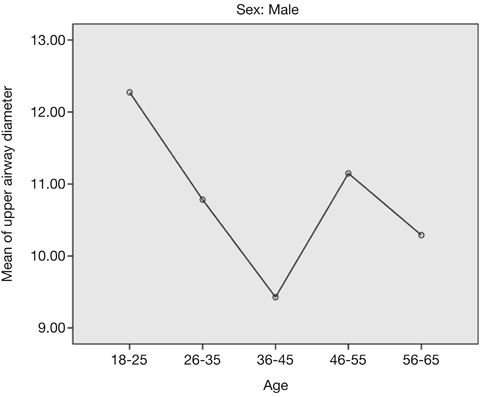
Fig. 2 Upper airway diameter in males according to age group.

Fig. 3 Upper airway diameter in females according to age group.
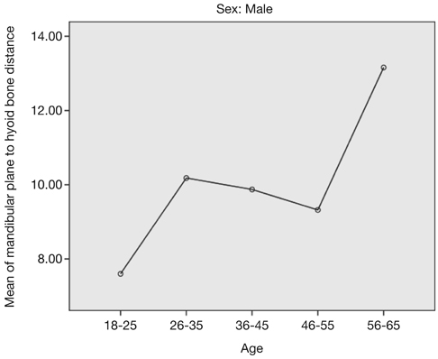
Fig. 4 Distance from the mandibular plane to the hyoid bone (MP-H) in males according to age group.

Fig. 5 Distance from the mandibular plane to the hyoid bone (MP-H) in females according to age group.
Reference
-
1. Fusetti M, Fioretti AB, Valenti M, Masedu F, Lauriello M, Pagliarella M. Cardiovascular and metabolic comorbidities in patients with obstructive sleep apnoea syndrome. Acta Otorhinolaryngol Ital. 2012; 32:320–325.2. Huang QR, Qin Z, Zhang S, Chow CM. Clinical patterns of obstructive sleep apnea and its comorbid conditions: a data mining approach. J Clin Sleep Med. 2008; 4:543–550.
Article3. Lavie P. Mortality in sleep apnoea syndrome: a review of the evidence. Eur Respir Rev. 2007; 16:203–210.
Article4. Grover M, Mookadam M, Armas D, Bozarth C, Castleberry T, Gannon M, et al. Identifying patients at risk for obstructive sleep apnea in a primary care practice. J Am Board Fam Med. 2011; 24:152–160.
Article5. Armalaite J, Lopatiene K. Lateral teleradiography of the head as a diagnostic tool used to predict obstructive sleep apnea. Dentomaxillofac Radiol. 2016; 45:20150085.
Article6. Sung MW, Lee WH, Wee JH, Lee CH, Kim E, Kim JW. Factors associated with hypertrophy of the lingual tonsils in adults with sleep-disordered breathing. JAMA Otolaryngol Head Neck Surg. 2013; 139:598–603.7. Friedel ME, Johnston DR, Singhal S, Al Khalili K, Farrell CJ, Evans JJ, et al. Airway management and perioperative concerns in acromegaly patients undergoing endoscopic transsphenoidal surgery for pituitary tumors. Otolaryngol Head Neck Surg. 2013; 149:840–844.
Article8. Sedaghat AR, Anderson IC, McGinley BM, Rossberg MI, Redett RJ, Ishman SL. Characterization of obstructive sleep apnea before and after tongue-lip adhesion in children with micrognathia. Cleft Palate Craniofac J. 2012; 49:21–26.
Article9. Pracharktam N, Nelson S, Hans MG, Broadbent BH, Redline S, Rosenberg C, et al. Cephalometric assessment in obstructive sleep apnea. Am J Orthod Dentofacial Orthop. 1996; 109:410–419.
Article10. Tangugsorn V, Skatvedt O, Krogstad O, Lyberg T. Obstructive sleep apnoea: a cephalometric study. Part I. Cervico-craniofacial skeletal morphology. Eur J Orthod. 1995; 17:45–56.
Article11. Szymanska J, Dobrowolska-Zarzycka M. The influence of upper airways diameter on the intensity of obstructive sleep apnea. Ann Agric Environ Med. 2014; 21:217–220.12. Martin SE, Mathur R, Marshall I, Douglas NJ. The effect of age, sex, obesity and posture on upper airway size. Eur Respir J. 1997; 10:2087–2090.
Article13. Gabbay IE, Lavie P. Age- and gender-related characteristics of obstructive sleep apnea. Sleep Breath. 2012; 16:453–460.
Article14. Guttal KS, Burde KN. Cephalometric evaluation of upper airway in healthy adult population: a preliminary study. J Oral Maxillofac Radiol. 2013; 1:55–60.
Article15. Block AJ, Boysen PG, Wynne JW, Hunt LA. Sleep apnea, hypopnea and oxygen desaturation in normal subjects. A strong male predominance. N Engl J Med. 1979; 300:513–517.16. Julià-Serdà G, Pérez-Peñate G, Saavedra-Santana P, Ponce-González M, Valencia-Gallardo JM, Rodríguez-Delgado R, et al. Usefulness of cephalometry in sparing polysomnography of patients with suspected obstructive sleep apnea. Sleep Breath. 2006; 10:181–187.
Article
- Full Text Links
-
- Actions
-
Cited
- CITED
-
- Close
- Share
-
- Similar articles
-
- Effects of Menopause on Obstructive Sleep Apnea
- Complications of Obstructive Sleep Apnea
- Role of oral and maxillofacial surgeons in the treatment of obstructive sleep apnea patients
- Predictors for Presence and Severity of Obstructive Sleep Apnea in Snoring Patients: Significance of Neck Circumference
- The Role of Endothelin-1 in Obstructive Sleep Apnea Syndrome and Pulmonary Hypertension
