J Korean Soc Spine Surg.
2017 Dec;24(4):241-245. 10.4184/jkss.2017.24.4.241.
Surfer's Myelopathy: A Case Report
- Affiliations
-
- 1Department of Orthopedic Surgery, Seoul St. Mary's Hospital, College of Medicine, The Catholic University of Korea, Seoul, Korea. boscoa@catholic.ac.kr
- KMID: 2408193
- DOI: http://doi.org/10.4184/jkss.2017.24.4.241
Abstract
- STUDY DESIGN: A case report.
OBJECTIVES
To report a rare cause of non-traumatic spinal cord injury (SCI) during surfing SUMMARY OF LITERATURE REVIEW: Surfer's myelopathy is a non-traumatic SCI associated with the hyperextension posture during paddling in surfing. Although the definite pathomechanism has not been identified, cord ischemia followed by arterial infarction may be related to this injury.
MATERIALS AND METHODS
A young healthy male patient presented with a SCI that occurred during his first time surfing. Magnetic resonance imaging revealed a T2-hyperintense lesion in the spinal cord from D10 to the conus medullaris.
RESULTS
The patient completely recovered without any neurologic deficits after steroid therapy and other forms of supportive management.
CONCLUSIONS
Since surfing is becoming more common in Korea, awareness of surfer's myelopathy is important for early diagnosis and proper management.
MeSH Terms
Figure
-
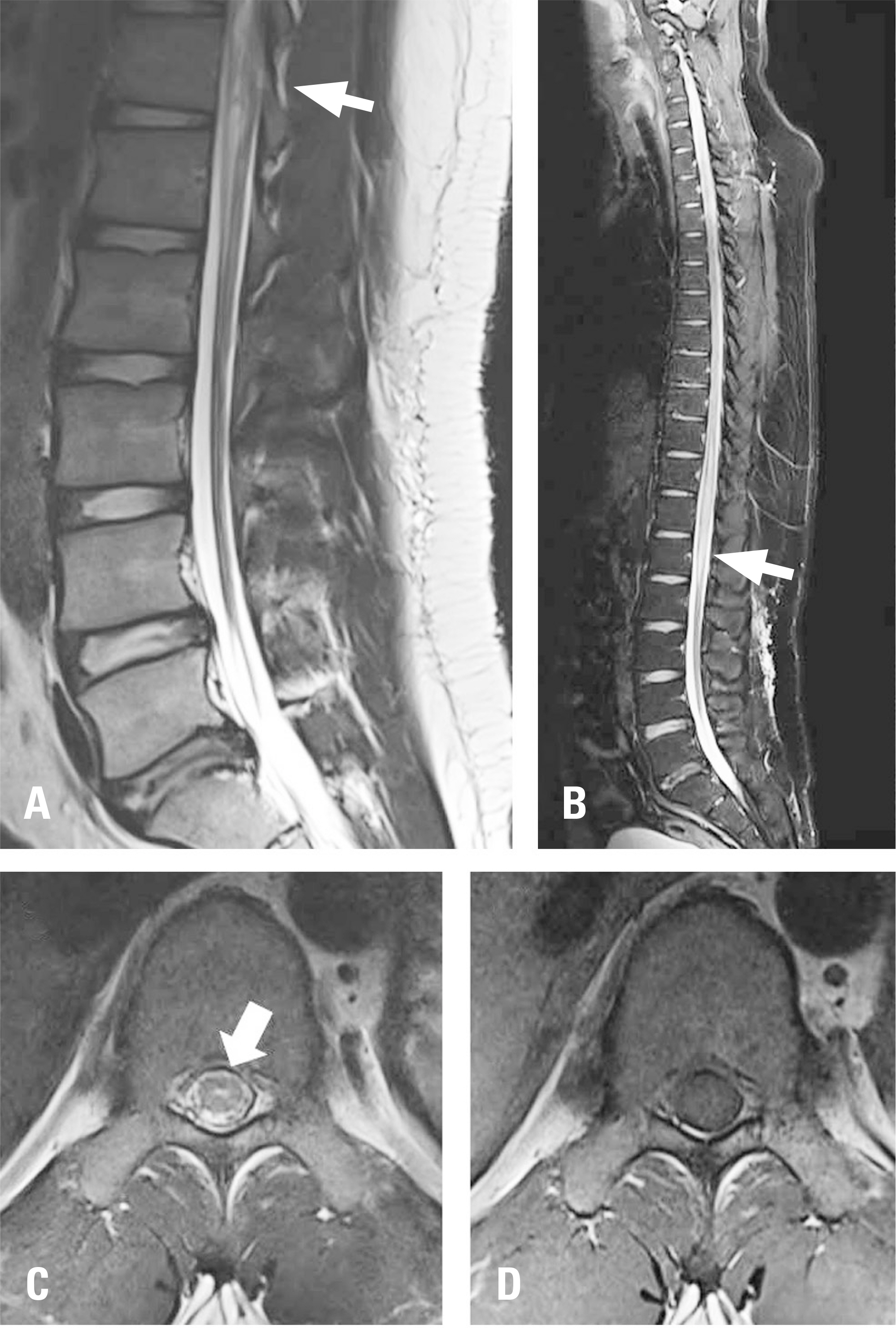
Fig. 1. Initial thoracolumbar spine magnetic resonance imaging (MRI) (A) and whole-spine MRI (B) showing a T2-hyperintense lesion extending from D10 to the conus medullaris (white arrow). On the D10 axial section, a T2-weighted image revealed a ‘pencil-like’ (white arrow) hyperintense central lesion (C), but normal signal intensity was observed on the T1-weighted image (D).
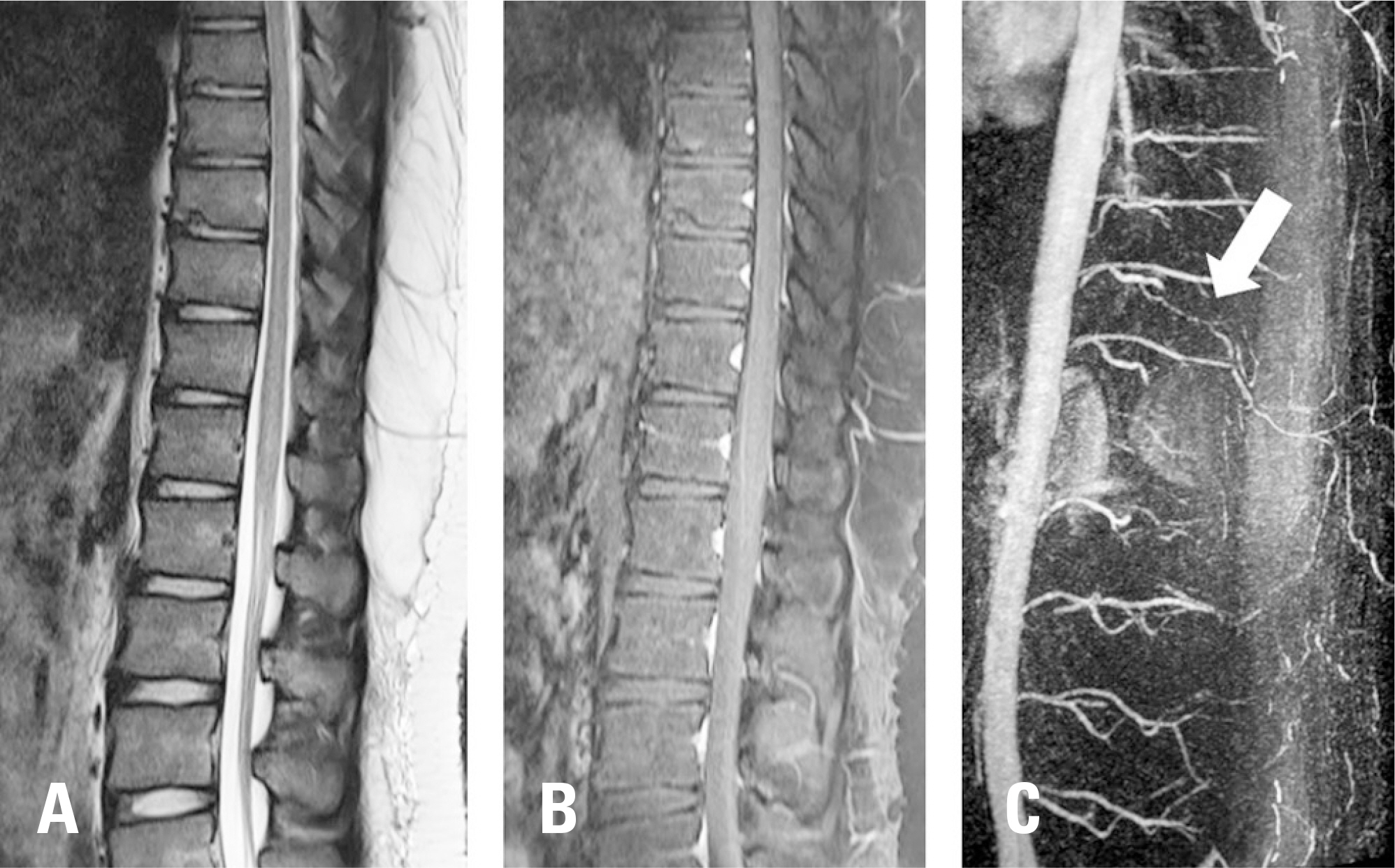
Fig. 2. A follow-up T2 weighted sagittal image (A) showing decreased signal intensity compared to the initial magnetic resonance imaging and normal signal intensity on a gadolinium-enhancing T1-weighted image (B). Magnetic resonance angiography revealing no occlusion or stenosis of any segmental artery or the Adamkiewicz artery (white arrow) (C).
Reference
-
1. Thompson TP, Pearce J, Chang G, et al. Surfer's myelopathy. Spine (Phila Pa 1976). 2004 Aug 15; 29(16):E353–6. DOI: 10.1097/01.BRS.0000134689.84162.E7.
Article2. Chang CW, Donovan DJ, Liem LK, et al. Surfers’ myelopathy: a case series of 19 novice surfers with nontraumatic myelopathy. Neurology. 2012 Nov 27; 79(22):2171–6. DOI: 10.1212/WNL.0b013e31827595cd.
Article3. Freedman BA, Malone DG, Rasmussen PA, et al. Surfer’'s Myelopathy: A Rare Form of Spinal Cord Infarction in Novice Surfers: A Systematic Review. Neurosurgery. 2016 May; 78(5):602–11. DOI: 10.1227/NEU.0000000000001089.4. Cheshire WP, Santos CC, Massey EW, et al. Spinal cord infarction: etiology and outcome. Neurology. 1996 Aug; 47(2):321–30. DOI: 10.1212/WNL.47.2.321.5. Baik MY, Jeong SH, Lee WW, et al. Surfer's myelopathy. J Korean Neurol Assoc. 2016; 34(2):145–9. DOI: 10.17340/jkna.2016.2.11.
Article6. White BC, Stirling AJ, Paterson E, et al. Diagnosis and management of patients at risk of or with metastatic spinal cord compression: summary of NICE guidance. BMJ. 2008 Nov 27; 337:a2538. DOI: 10.1136/bmj.a2538.
Article7. Nakamoto BK, Siu AM, Hashiba KA, et al. Surfer's Myelopathy: a radiologic study of 23 cases. AJNR Am J Neuroradiol. 2013 Dec; 34(12):2393–8. DOI: 10.3174/ajnr. A3599.
Article8. Kudo K, Terae S, Asano T, et al. Anterior spinal artery and artery of Adamkiewicz detected by using multi-detector row CT. AJNR Am J Neuroradiol. 2003 Jan; 24(1):13–7.9. Tanaka H, Minatoya K, Matsuda H, et al. Embolism is emerging as a major cause of spinal cord injury after descending and thoracoabdominal aortic repair with a contemporary approach: magnetic resonance findings of spinal cord injury. Interact Cardiovasc Thorac Surg. 2014 Aug; 19(2):205–10. DOI: 10.1093/icvts/ivu148.
Article10. Lieske J, Cameron B, Drinkwine B, et al. Surfer's myelopathy-demonstrated by diffusion-weighted magnetic resonance imaging: a case report and literature review. J Comput Assist Tomogr. 2011 Jul-Aug; 35(4):492–4. DOI: 10.1097/RCT.0b013e31821e277b.
