Recurrent Acute Myocardial Infarction Caused by Intra-cardiac Metastatic Undifferentiated Pleomorphic Sarcoma during Cancer Treatment
- Affiliations
-
- 1Division of Cardiovascular Medicine, Department of Internal Medicine, Dankook University College of Medicine, Dankook University Hospital, Cheonan, Korea. neosoo70@dankook.ac.kr
- 2Department of Pathology, Dankook University College of Medicine, Cheonan, Korea.
- KMID: 2407680
- DOI: http://doi.org/10.4250/jcu.2018.26.1.40
Abstract
- No abstract available.
Keyword
Figure
-
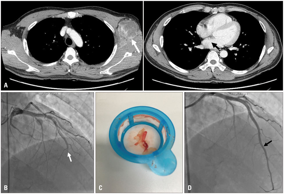
Fig. 1 Computed tomography and coronary angiography. A: Initial chest computed tomography showed a large, infiltrative, and heterogeneously soft tissue mass at the left axilla (white arrow). There was no definite mass in the left atrium (black arrow). B: Coronary angiography showed the total occlusion of the distal left anterior descending artery (white arrow). C: The aspirated tissue material appeared as mucoid and whitish debris. D: The final coronary angiography showed that flow had been completely restored (black arrow).
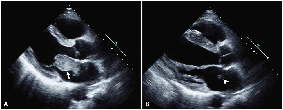
Fig. 2 Echocardiography. A: Echocardiography showed a huge mass with heterogeneous echogenicity in the left atrium affecting through the mitral valve leaflet before six months ago, at the time of pembrolizumab treatment (white arrow). B: The left atrium tumor mass was significantly decreased at the time of admission in the echocardiography (white arrow head).
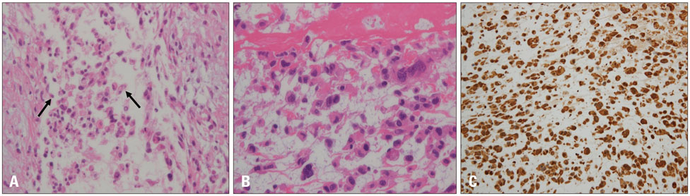
Fig. 3 Pathology findings. A: The primary axillary tumor was cytologically compatible with undifferentiated pleomorphic sarcoma that focally showed discohesive and relatively monotonous round cell components (black arrows) (H&E, × 200). B: The coronary embolus tissues showed similar round cell morphology and a few scattered pleomorphic giant cells (H&E, × 400). C: The tumor cells were diffusely immunoreactive with vimentin, a representative mesenchymal marker (ABC, × 200). H&E: hematoxylin and eosin.
Reference
-
1. Hallahan DE, Vogelzang NJ, Borow KM, Bostwick DG, Simon MA. Cardiac metastases from soft-tissue sarcomas. J Clin Oncol. 1986; 4:1662–1669.
Article2. Takenaka S, Hashimoto N, Araki N, Hamada K, Naka N, Joyama S, Kakunaga S, Ueda T, Myoui A, Yoshikawa H. Eleven cases of cardiac metastases from soft-tissue sarcomas. Jpn J Clin Oncol. 2011; 41:514–518.
Article3. Ali MK, Ewer MS, Cangir A, Fisher DJ. Coronary artery embolism following cancer chemotherapy. Am J Pediatr Hematol Oncol. 1987; 9:200–203.4. Chinen K, Kurosumi M, Ohkura Y, Sakamoto A, Fujioka Y. Sudden unexpected death in patients with malignancy: a clinicopathologic study of 28 autopsy cases. Pathol Res Pract. 2006; 202:869–875.
Article5. Steiner I, Vojáček J. Carcinoma embolization in coronary artery causing myocardial infarction: diagnosis from coronary thromboaspirate. Pathol Res Pract. 2014; 210:198–200.
- Full Text Links
-
- Actions
-
Cited
- CITED
-
- Close
- Share
-
- Similar articles
-
- Cutaneous Metastatic Undifferentiated Pleomorphic Sarcoma from a Mediastinal Sarcoma
- A Case of Pleomorphic Dermal Sarcoma Showing Characteristics of Myxoinflammatory Fibroblastic Sarcoma
- Primary Undifferentiated Pleomorphic Sarcoma of the Left Atrium that Presented as Acute Pulmonary Edema
- Undifferentiated Pleomorphic Sarcoma of the Male Breast Causing Diagnostic Challenges
- Sinonasal Undifferentiated Pleomorphic Sarcoma in Five Patient Cases
