Korean J Orthod.
2018 Mar;48(2):98-106. 10.4041/kjod.2018.48.2.98.
Adjunctive buccal and palatal corticotomy for adult maxillary expansion in an animal model
- Affiliations
-
- 1Department of Paediatric Dentistry and Orthodontics, Faculty of Dentistry, University of Malaya, Kuala Lumpur, Malaysia. zamrir@um.edu.my
- 2Department of Veterinary Clinical Studies, Faculty of Veterinary Medicine, Universiti Putra Malaysia, Selangor, Malaysia.
- 3Department of Diagnostic and Intergrated Dental Practice, Faculty of Dentistry, University of Malaya, Kuala Lumpur, Malaysia.
- 4Department of Restorative Dentistry, Faculty of Dentistry, University of Malaya, Kuala Lumpur, Malaysia.
- KMID: 2406818
- DOI: http://doi.org/10.4041/kjod.2018.48.2.98
Abstract
OBJECTIVE
This study aimed to explore the usefulness of adjunctive buccal and palatal corticotomy for adult maxillary expansion in an animal model using cone-beam computed tomography (CBCT).
METHODS
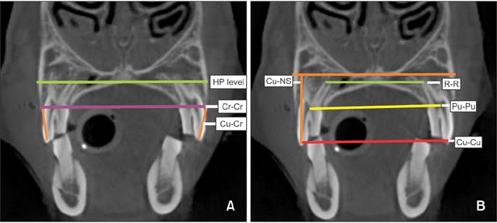
Twelve adult sheep were randomly divided into two groups (each n = 6): a control group, where no treatment was administered, and a treatment group, where buccal and palatal corticotomy-assisted maxillary expansion was performed. CBCT scans were taken before (T1) and after (T2) treatment. Differences in all transverse dental and alveolar dimensions, alveolar width at crest level, hard palate level, horizontal bone loss, interdental cusp width and inter-root apex were assessed using Wilcoxon signed-rank and Mann-Whitney U-tests. Kruskal-Wallis tests and pairwise comparisons were used to detect the significance of differences among the inter-premolar and inter-molar widths.
RESULTS
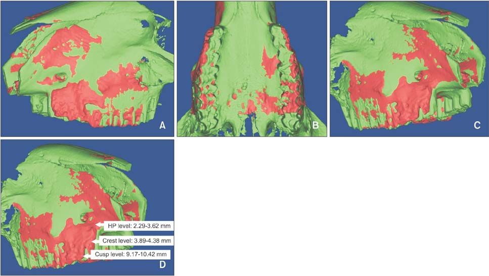
CBCT data revealed significant changes in all transverse dental and alveolar dimensions. The mean interpremolar alveolar width showed an increase of 2.29 to 3.62 mm at the hard palate level, 3.89 to 4.38 mm at the alveolar crest level, and 9.17 to 10.42 mm at the buccal cusp level. Dental changes in the vertical dimension were not significant.
CONCLUSIONS
Our findings based on an adult animal model suggest that adjunctive buccal and palatal corticotomy can allow for both skeletal and dental expansion, with the amount of dental expansion exceeding that of skeletal expansion at alveolar crest and hard palate levels by two and three folds, respectively. Therefore, this treatment modality is potential to enhance the outcomes of maxillary expansion in adults.
Keyword
MeSH Terms
Figure
Cited by 2 articles
-
Corticotomy for orthodontic tooth movement
Won Lee
J Korean Assoc Oral Maxillofac Surg. 2018;44(6):251-258. doi: 10.5125/jkaoms.2018.44.6.251.Alveolar restoration following rapid maxillary expansion with and without corticotomy: A microcomputed tomography study in sheep
My Huy Thuc Le, Abu Kasim Noor Hayaty, Zuraiza Mohamad Zaini, Sulaiman Md Dom, Norliza Ibrahim, Zamri Bin Radzi
Korean J Orthod. 2019;49(4):235-245. doi: 10.4041/kjod.2019.49.4.235.
Reference
-
1. Northway W. Palatal expansion in adults: the surgical approach. Am J Orthod Dentofacial Orthop. 2011; 140:463. 465. 467 passim.
Article2. Handelman C. Palatal expansion in adults: the nonsurgical approach. Am J Orthod Dentofacial Orthop. 2011; 140:462. 464. 466 passim.
Article3. McNamara JA Jr, Baccetti T, Franchi L, Herberger TA. Rapid maxillary expansion followed by fixed appliances: a long-term evaluation of changes in arch dimensions. Angle Orthod. 2003; 73:344–353.4. Gurel HG, Memili B, Erkan M, Sukurica Y. Long-term effects of rapid maxillary expansion followed by fixed appliances. Angle Orthod. 2010; 80:5–9.
Article5. Lagravere MO, Major PW, Flores-Mir C. Long-term skeletal changes with rapid maxillary expansion: a systematic review. Angle Orthod. 2005; 75:1046–1052.6. Suri L, Taneja P. Surgically assisted rapid palatal expansion: a literature review. Am J Orthod Dentofacial Orthop. 2008; 133:290–302.
Article7. Handelman CS, Wang L, BeGole EA, Haas AJ. Nonsurgical rapid maxillary expansion in adults: report on 47 cases using the Haas expander. Angle Orthod. 2000; 70:129–144.8. Bishara SE, Staley RN. Maxillary expansion: clinical implications. Am J Orthod Dentofacial Orthop. 1987; 91:3–14.
Article9. Bassarelli T, Dalstra M, Melsen B. Changes in clinical crown height as a result of transverse expansion of the maxilla in adults. Eur J Orthod. 2005; 27:121–128.
Article10. Angelieri F, Cevidanes LH, Franchi L, Gonçalves JR, Benavides E, McNamara JA Jr. Midpalatal suture maturation: classification method for individual assessment before rapid maxillary expansion. Am J Orthod Dentofacial Orthop. 2013; 144:759–769.
Article11. Knaup B, Yildizhan F, Wehrbein H. Age-related changes in the midpalatal suture. A histomorphometric study. J Orofac Orthop. 2004; 65:467–474.12. Korbmacher H, Schilling A, Püschel K, Amling M, Kahl-Nieke B. Age-dependent three-dimensional microcomputed tomography analysis of the human midpalatal suture. J Orofac Orthop. 2007; 68:364–376.
Article13. Kole H. Surgical operations on the alveolar ridge to correct occlusal abnormalities. Oral Surg Oral Med Oral Pathol. 1959; 12:515–529.
Article14. Choo H, Heo HA, Yoon HJ, Chung KR, Kim SH. Treatment outcome analysis of speedy surgical orthodontics for adults with maxillary protrusion. Am J Orthod Dentofacial Orthop. 2011; 140:e251–e262.
Article15. Chung KR, Kim SH, Lee BS. Speedy surgical-orthodontic treatment with temporary anchorage devices as an alternative to orthognathic surgery. Am J Orthod Dentofacial Orthop. 2009; 135:787–798.
Article16. Chung KR, Mitsugi M, Lee BS, Kanno T, Lee W, Kim SH. Speedy surgical orthodontic treatment with skeletal anchorage in adults--sagittal correction and open bite correction. J Oral Maxillofac Surg. 2009; 67:2130–2148.
Article17. Lines PA. Adult rapid maxillary expansion with corticotomy. Am J Orthod. 1975; 67:44–56.
Article18. Echchadi ME, Benchikh B, Bellamine M, Kim SH. Corticotomy-assisted rapid maxillary expansion: A novel approach with a 3-year follow-up. Am J Orthod Dentofacial Orthop. 2015; 148:138–153.
Article19. Charan J, Kantharia ND. How to calculate sample size in animal studies? J Pharmacol Pharmacother. 2013; 4:303–306.
Article20. Wertz RA. Skeletal and dental changes accompanying rapid midpalatal suture opening. Am J Orthod. 1970; 58:41–66.
Article21. Cao Y, Zhou Y, Song Y, Vanarsdall RL Jr. Cephalometric study of slow maxillary expansion in adults. Am J Orthod Dentofacial Orthop. 2009; 136:348–354.
Article22. Lin L, Ahn HW, Kim SJ, Moon SC, Kim SH, Nelson G. Tooth-borne vs bone-borne rapid maxillary expanders in late adolescence. Angle Orthod. 2015; 85:253–262.
Article23. Lagravère MO, Major PW, Flores-Mir C. Dental and skeletal changes following surgically assisted rapid maxillary expansion. Int J Oral Maxillofac Surg. 2006; 35:481–487.
Article24. O'Ryan F, Schendel S. Nasal anatomy and maxillary surgery. II. Unfavorable nasolabial esthetics following the Le Fort I osteotomy. Int J Adult Orthodon Orthognath Surg. 1989; 4:75–84.25. Brunetto M, Andriani Jda S, Ribeiro GL, Locks A, Correa M, Correa LR. Three-dimensional assessment of buccal alveolar bone after rapid and slow maxillary expansion: a clinical trial study. Am J Orthod Dentofacial Orthop. 2013; 143:633–644.
Article26. Rungcharassaeng K, Caruso JM, Kan JY, Kim J, Taylor G. Factors affecting buccal bone changes of maxillary posterior teeth after rapid maxillary expansion. Am J Orthod Dentofacial Orthop. 2007; 132:428.e1–428.e8.
Article27. Gauthier C, Voyer R, Paquette M, Rompré P, Papadakis A. Periodontal effects of surgically assisted rapid palatal expansion evaluated clinically and with cone-beam computerized tomography: 6-month preliminary results. Am J Orthod Dentofacial Orthop. 2011; 139:4 Suppl. S117–S128.
Article28. Forst D, Nijjar S, Khaled Y, Lagravere M, Flores-Mir C. Radiographic assessment of external root resorption associated with jackscrew-based maxillary expansion therapies: a systematic review. Eur J Orthod. 2014; 36:576–585.
Article29. Robiony M, Polini F, Costa F, Zerman N, Politi M. Ultrasonic bone cutting for surgically assisted rapid maxillary expansion (SARME) under local anaesthesia. Int J Oral Maxillofac Surg. 2007; 36:267–269.
Article
- Full Text Links
- Actions
-
Cited
- CITED
-
- Close
- Share
- Similar articles
-
- A posteroanterior cephalometric study on the change of maxilla by rapid palatal expansion
- Photoelastic evaluation of maxillary posterior crossbite appliance
- Crowding with no posterior crossbite treatment byrapid palatal expansion
- A cone-beam computed tomography evaluation of buccal bone thickness following maxillary expansion
- A study on the effect of rapid maxillary expansion and its relapse


