Where Is the Ulnar Styloid Process? Identification of the Absolute Location of the Ulnar Styloid Process Based on CT and Verification of Neutral Forearm Rotation on Lateral Radiographs of the Wrist
- Affiliations
-
- 1Department of Orthopedic Surgery, Seoul St. Mary's Hospital, College of Medicine, The Catholic University of Korea, Seoul, Korea. ygchung@catholic.ac.kr
- 2Department of Radiology, Seoul St. Mary's Hospital, College of Medicine, The Catholic University of Korea, Seoul, Korea.
- KMID: 2405486
- DOI: http://doi.org/10.4055/cios.2018.10.1.80
Abstract
- BACKGROUND
The location of the ulnar styloid process can be confusing because the radius and the hand rotate around the ulna. The purpose of this study was to identify the absolute location of the ulnar styloid process, which is independent of forearm pronation or supination, to use it as a reference for neutral forearm rotation on lateral radiographs of the wrist.
METHODS
Computed tomography (CT) images of 23 forearms taken with elbow flexion of 70° to 90° were analyzed. The axial CT images were reconstructed to be perpendicular to the distal ulnar shaft. The absolute location of the ulnar styloid process in this study was defined as the position of the ulnar styloid process on the axial plane of the ulnar head relative to the long axis of the humeral shaft with the elbow set in the position for standard lateral radiographs of the wrist. To identify in which direction the ulnar styloid is located on the axial plane of the ulnar head, the angle between "the line of humeral long axis projected on the axial plane of the ulna" and "the line passing the center of the ulnar head and the center of the ulnar styloid" was measured (ulnar styloid direction angle). To identify how volarly or dorsally the ulnar styloid should appear on the true lateral view of the wrist, the ratio of "the volar-dorsal diameter of the ulnar head" and "the distance between the volar-most aspect of the ulnar head and the center of the ulnar styloid" was calculated (ulnar styloid location ratio).
RESULTS
The mean ulnar styloid direction angle was 12° dorsally. The mean ulnar styloid location ratio was 1:0.55.
CONCLUSIONS
The ulnar styloid is located at nearly the ulnar-most (the opposite side of the humerus with the elbow flexed) and slightly dorsal aspects of the ulnar head on the axial plane. It should appear almost midway (55% dorsally) from the ulnar head on the standard lateral view of the wrist in neutral forearm rotation. These location references could help clinicians determine whether the forearm is in neutral or rotated position on an axial CT/magnetic resonance imaging scan or a lateral radiograph of the wrist.
Keyword
Figure
-
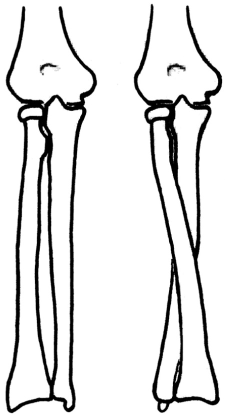
Fig. 1 An example of illustrations of forearm pronation and supination. Note the ulnar styloid process is drawn at different locations of the distal ulna in the two pictures although the proximal ulna is in the same position.
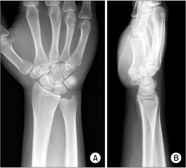
Fig. 2 Posteroanterior and lateral radiographs of the wrist commonly seen in practice. Note the ulnar styloid process is in the same location on the posteroanterior (A) and lateral (B) views. This is not realistic assuming that the two radiographs are taken from the same neutrally-rotated wrist with X-ray beams angled at 90°.
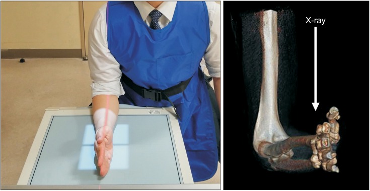
Fig. 3 Positioning for standard lateral radiographs of the wrist. The elbow is flexed to 90°, the arm is adducted against the trunk, and the forearm is on the film plate in neutral rotation. The X-ray beam is aligned vertically over the radial styloid 100 cm from the film. In this position, the central beam is almost parallel to the long axis of the humerus.
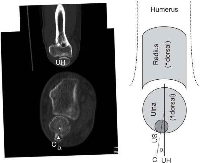
Fig. 4 Measurement of ulnar styloid (US) direction angle. A straight line passing through the center of the ulnar head and parallel to the long axis of the humerus (line UH) was drawn on the axial image of the wrist computed tomography obtained with the elbow flexed (arrowhead: center of the ulnar styloid process, asterisk: center of the ulnar head). Then a straight line was drawn from the center of the ulnar head to the center of the ulnar styloid process (line C). To identify in which direction the ulnar styloid process was located on the axial plane of the ulnar head, the angle between line C and line UH (US direction angle = angle α) was measured.
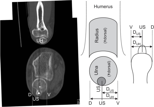
Fig. 5 Calculation of ulnar styloid (US) location ratio. Straight lines passing through the most volar and dorsal points of the ulnar head (line V and line D) and the center of the ulnar styloid process (line US) parallel to the long axis of the humerus (line UH) were drawn on the axial image of the wrist computed tomography obtained with the elbow flexed (arrowhead: center of the ulnar styloid process, asterisk: center of the ulnar head). To identify how volarly or dorsally the ulnar styloid process should be located on the lateral view, the ratio of the distance between line V and line D (DUH, diameter of the ulnar head) and the distance between line V and line US (DUS, distance of the US) was calculated (US location ratio = DUS/DUH).
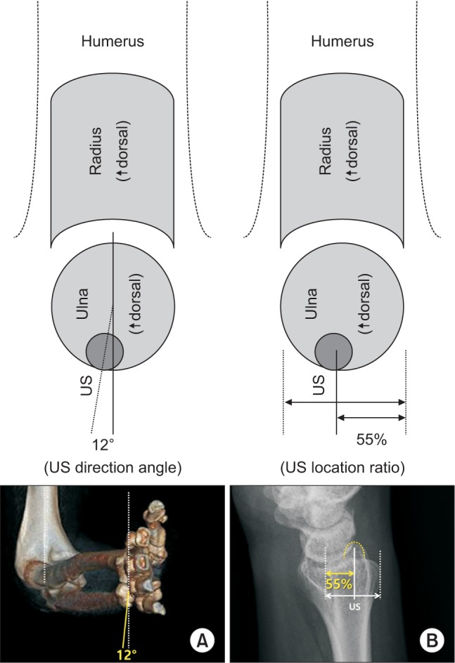
Fig. 6 The absolute location of the ulnar styloid process on the axial plane of the ulnar head relative to the long axis of the humeral shaft with the elbow set in the position for standard wrist lateral radiographs. (A) The mean ulnar styloid (US) direction angle is 12° ± 13° dorsally. (B) The mean US location ratio is 1 : 0.55 ± 0.09.
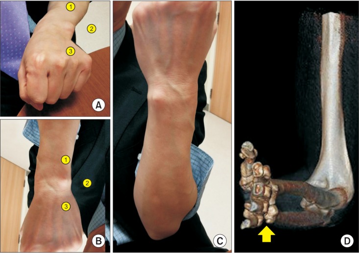
Fig. 7 The location of the ulnar styloid process relative to the hand. Where is the ulnar styloid in (A) and (B)? Although its position may be thought to be similar in (A) and (B), the actual location of it is different. In fact, (A) shows a pronated wrist and (B) shows a supinated wrist. (B) is a 180°-rotated image of the original photo (C). Therefore, the closest answer is number ③ in (A) and number ① in (B). (D) The ulnar styloid process is located on the opposite side of the humerus when the elbow is flexed regardless of the forearm rotation (arrow: ulnar styloid process).

Fig. 8 Locations of the ulnar styloid process in the wrist with the forearm pronated (A, C) and supinated (B, D). The ulnar styloid process may be located either volar (A, C) or dorsal (B, D) to the hand when the forearm is pronated or supinated.
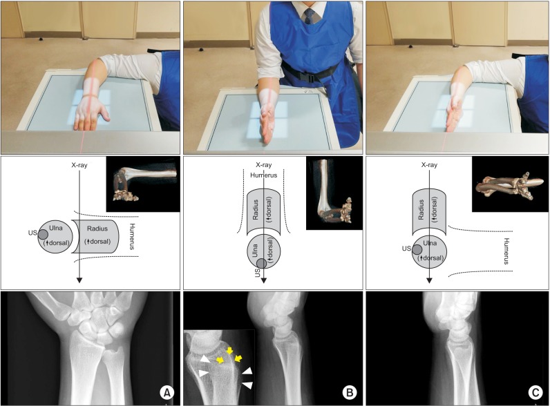
Fig. 9 Forearm rotation and relevant wrist radiographs. (A) Standard posteroanterior view with neutral forearm rotation. (B) Standard lateral view with neutral forearm rotation (arrows: ulnar styloid process, arrowheads: ulnar head). Note the ulnar styloid process is located almost midway from the ulnar head. (C) Lateral view taken with the forearm supinated. Note the distal ulna looks almost the same as that on the posteroanterior view. US: ulnar styloid.
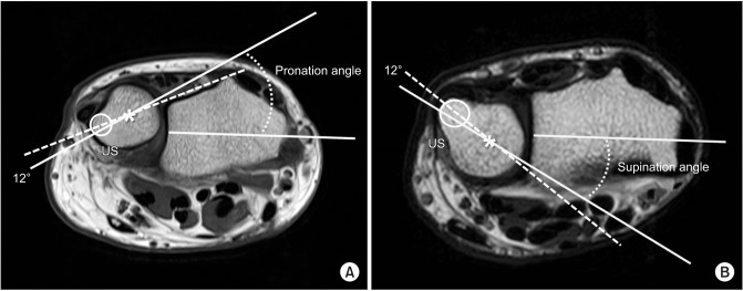
Fig. 10 Measurement of forearm rotation on axial images of wrist magnetic resonance imaging (MRI). It is possible to determine the amount of forearm pronation (A) and supination (B) on existing axial MRI or computed tomography images of the wrist by referencing the direction of the ulnar styloid (US) relative to the radius (asterisks: center of the ulnar head, circle: US).
Cited by 1 articles
-
Ultrasonographic Analysis of Optimal Needle Placement for Extensor Indicis
Jin Young Kim, Hyun Seok, Sang-Hyun Kim, Yoon-Hee Choi, Jun Young Ahn, Seung Yeol Lee
Ann Rehabil Med. 2020;44(6):450-458. doi: 10.5535/arm.20035.
Reference
-
1. Quigley RJ, Robicheaux GW, Lee TQ. The proximal and distal position of the radius relative to the ulna through a full range of elbow flexion and forearm rotation. J Hand Surg Eur Vol. 2014; 39(5):535–540. PMID: 23695153.
Article2. Tomaino MM. The importance of the pronated grip x-ray view in evaluating ulnar variance. J Hand Surg Am. 2000; 25(2):352–357. PMID: 10722828.
Article3. Parker AS, Nguyen M, Minard CG, Guffey D, Willis MH, Reichel LM. Measurement of ulnar variance from the lateral radiograph: a comparison of techniques. J Hand Surg Am. 2014; 39(6):1114–1121. PMID: 24810937.
Article4. Nakamura R, Horii E, Imaeda T, Tsunoda K, Nakao E. Distal radioulnar joint subluxation and dislocation diagnosed by standard roentgenography. Skeletal Radiol. 1995; 24(2):91–94. PMID: 7747190.
Article5. Schreibman KL, Freeland A, Gilula LA, Yin Y. Imaging of the hand and wrist. Orthop Clin North Am. 1997; 28(4):537–582. PMID: 9257964.
Article6. Bhat AK, Kumar B, Acharya A. Radiographic imaging of the wrist. Indian J Plast Surg. 2011; 44(2):186–196. PMID: 22022028.
Article7. Hagert E, Hagert CG. Understanding stability of the distal radioulnar joint through an understanding of its anatomy. Hand Clin. 2010; 26(4):459–466. PMID: 20951895.
Article8. Loredo RA, Sorge DG, Garcia G. Radiographic evaluation of the wrist: a vanishing art. Semin Roentgenol. 2005; 40(3):248–289. PMID: 16060116.
Article9. Vezeridis PS, Yoshioka H, Han R, Blazar P. Ulnar-sided wrist pain. Part I: anatomy and physical examination. Skeletal Radiol. 2010; 39(8):733–745. PMID: 19722104.
Article10. Campbell D, Campbell R, O'Connor P, Hawkes R. Sportsrelated extensor carpi ulnaris pathology: a review of functional anatomy, sports injury and management. Br J Sports Med. 2013; 47(17):1105–1111. PMID: 24096897.
Article11. Ehman EC, Hayes ML, Berger RA, Felmlee JP, Amrami KK. Subluxation of the distal radioulnar joint as a predictor of foveal triangular fibrocartilage complex tears. J Hand Surg Am. 2011; 36(11):1780–1784. PMID: 22036278.
Article12. Darcus HD, Salter N. The amplitude of pronation and supination with the elbow flexed to a right angle. J Anat. 1953; 87(2):169–184. PMID: 13044728.13. Palmer AK, Glisson RR, Werner FW. Ulnar variance determination. J Hand Surg Am. 1982; 7(4):376–379. PMID: 7119397.
Article14. Duck TR, Dunning CE, Armstrong AD, Johnson JA, King GJ. Application of screw displacement axes to quantify elbow instability. Clin Biomech (Bristol, Avon). 2003; 18(4):303–310.
Article
- Full Text Links
-
- Actions
-
Cited
- CITED
-
- Close
- Share
-
- Similar articles
-
- Operative Treatment for Ulnar Styloid Process Fractures with Unstable Intraarticular Distal Radius Fractures
- The 'beta-wire Technique' for the Fixation of Ulnar Styloid Process Fracture: Surgical Technique
- Treatment of Ulnar Fractures Combined with Distal Radius Fracture
- A study on the styloid process in panoramic radiographs
- Anatomic Characteristics of Pronator Quadratus Muscle: A Cadaver Study
