Evaluation of the Subscapularis Tendon Tears on 3T Magnetic Resonance Arthrography: Comparison of Diagnostic Performance of T1-Weighted Spectral Presaturation with Inversion-Recovery and T2-Weighted Turbo Spin-Echo Sequences
- Affiliations
-
- 1Department of Radiology, Kyungpook National University Hospital, Daegu 41944, Korea.
- 2Department of Radiology, Seoul National University Bundang Hospital, Seongnam 13620, Korea. joongmoahn@gmail.com
- 3Department of Orthopaedic Surgery, Seoul National University Bundang Hospital, Seongnam 13620, Korea.
- KMID: 2404929
- DOI: http://doi.org/10.3348/kjr.2018.19.2.320
Abstract
OBJECTIVE
To compare the T1-weighted spectral presaturation with inversion-recovery sequences (T1 SPIR) with T2-weighted turbo spin-echo sequences (T2 TSE) on 3T magnetic resonance arthrography (MRA) in the evaluation of the subscapularis (SSC) tendon tear with arthroscopic findings as the reference standard.
MATERIALS AND METHODS
This retrospective study included 120 consecutive patients who had undergone MRA within 3 months between April and December 2015. Two musculoskeletal radiologists blinded to the arthroscopic results evaluated T1 SPIR and T2 TSE images in separate sessions for the integrity of the SSC tendon, examining normal/articular-surface partial-thickness tear (PTTa)/full-thickness tear (FTT). Diagnostic performance of T1 SPIR and T2 TSE was calculated with arthroscopic results as the reference standard, and sensitivity, specificity, and accuracy were compared using the McNemar test. Interobserver agreement was measured with kappa (κ) statistics.
RESULTS
There were 74 SSC tendon tears (36 PTTa and 38 FTT) confirmed by arthroscopy. Significant differences were found in the sensitivity and accuracy between T1 SPIR and T2 TSE using the McNemar test, with respective rates of 95.9-94.6% vs. 71.6-75.7% and 90.8-91.7% vs. 79.2-83.3% for detecting tear; 55.3% vs. 31.6-34.2% and 85.8% vs. 78.3-79.2%, respectively, for FTT; and 91.7-97.2% vs. 58.3-61.1% and 89% vs. 78-79.3%, respectively, for PTTa. Interobserver agreement for T1 SPIR was almost perfect for T1 SPIR (κ = 0.839) and substantial for T2 TSE (κ = 0.769).
CONCLUSION
T1-weighted spectral presaturation with inversion-recovery sequences is more sensitive and accurate compared to T2 TSE in detecting SSC tendon tear on 3T MRA.
Keyword
MeSH Terms
Figure
-
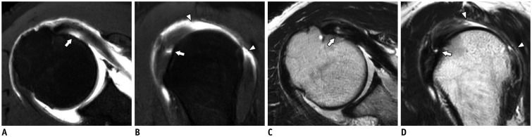
Fig. 1 67-year-old woman with arthroscopically confirmed articular-sided partial-thickness SSC tendon tear.T1-weighted spectral presaturation with inversion-recovery axial (A) and oblique sagittal (B) images and T2-weighted turbo spin-echo axial (C) and oblique sagittal (D) images demonstrate localized contrast leakage onto uppermost facet of lesser tuberosity (arrows). Large contrast-filled defect caused by complete tear of supraspinatus and infraspinatus tendons (arrowheads) is also noted. SSC = subscapularis
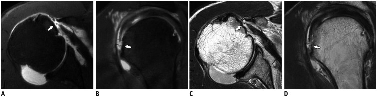
Fig. 2 64-year-old man with full-thickness SSC tendon tear diagnosed by arthroscopy.T1-weighted spectral presaturation with inversion-recovery axial (A) and oblique sagittal (B) images and T2-weighted turbo spin-echo axial (C) and oblique sagittal (D) images reveal discontinuity of the tendon (arrows) with medial retraction.
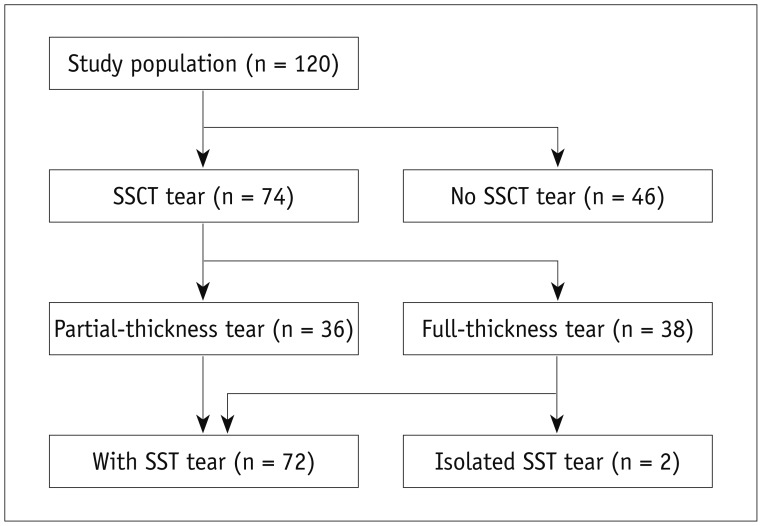
Fig. 3 Flowchart of arthroscopic results.SSCT = subscapularis tendon, SST = supraspinatus tendon
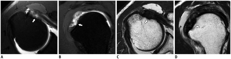
Fig. 4 68-year-old man with articular-sided partial-thickness SSC tendon tear confirmed by arthroscopy.A, B. T1-weighted spectral presaturation with inversion-recovery axial (A) and oblique sagittal (B) images show focal contrast-filled defect in undersurface of uppermost footprint of SSC tendon (arrows), which was diagnosed as partial-thickness tear by both readers. C, D. T2-weighted turbo spin-echo axial (C) and oblique sagittal (D) images show slightly increased signal intensity of SSC tendon without fluid-equivalant signal defect (arrows), which was underdiagnosed as normal by both readers.
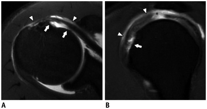
Fig. 5 68-year-old man with arthroscopically diagnosed full-thickness SSC tendon tear.A, B. T1-weighted spectral presaturation with inversion-recovery axial (A) and oblique sagittal (B) images show detachment of SSC tendon from lesser tuberosity with contrast filling (arrows). Superficial “bridging fiber” remains without retraction, which have continuity with fibers overlying bicipital groove and anterior-most fibers of supraspinatus tendon (arrowheads). Both readers underdiagnosed as partial-thickness SSC tear. Accompanied full-thickness supraspinatus tendon tear (asterisk) with contrast leakage into subacromial-subdeltoid bursal space is also shown.
Reference
-
1. Blasier RB, Soslowsky LJ, Malicky DM, Palmer ML. Posterior glenohumeral subluxation: active and passive stabilization in a biomechanical model. J Bone Joint Surg Am. 1997; 79:433–440. PMID: 9070535.2. Halder A, Zobitz ME, Schultz E, An KN. Structural properties of the subscapularis tendon. J Orthop Res. 2000; 18:829–834. PMID: 11117307.
Article3. Abboud JA, Soslowsky LJ. Interplay of the static and dynamic restraints in glenohumeral instability. Clin Orthop Relat Res. 2002; 48–57. PMID: 12072745.
Article4. Morag Y, Jamadar DA, Miller B, Dong Q, Jacobson JA. The subscapularis: anatomy, injury, and imaging. Skeletal Radiol. 2011; 40:255–269. PMID: 20033149.
Article5. Bergin D, Parker L, Zoga A, Morrison W. Abnormalities on MRI of the subscapularis tendon in the presence of a full-thickness supraspinatus tendon tear. AJR Am J Roentgenol. 2006; 186:454–459. PMID: 16423952.
Article6. Sakurai G, Ozaki J, Tomita Y, Kondo T, Tamai S. Incomplete tears of the subscapularis tendon associated with tears of the supraspinatus tendon: cadaveric and clinical studies. J Shoulder Elbow Surg. 1998; 7:510–515. PMID: 9814932.
Article7. MacMahon PJ, Taylor DH, Duke D, Brennan DD, O'Brien J, Eustace SJ. Contribution of full-thickness supraspinatus tendon tears to acquired subcoracoid impingement. Clin Radiol. 2007; 62:556–563. PMID: 17467393.
Article8. Burkhart SS. Arthroscopic treatment of massive rotator cuff tears. Clinical results and biomechanical rationale. Clin Orthop Relat Res. 1991; 45–56.9. Aluisio FV, Osbahr DC, Speer KP. Analysis of rotator cuff muscles in adult human cadaveric specimens. Am J Orthop (Belle Mead NJ). 2003; 32:124–129. PMID: 12647876.10. Foad A, Wijdicks CA. The accuracy of magnetic resonance imaging and magnetic resonance arthrogram versus arthroscopy in the diagnosis of subscapularis tendon injury. Arthroscopy. 2012; 28:636–641. PMID: 22281195.
Article11. Adams CR, Schoolfield JD, Burkhart SS. Accuracy of preoperative magnetic resonance imaging in predicting a subscapularis tendon tear based on arthroscopy. Arthroscopy. 2010; 26:1427–1433. PMID: 20875724.
Article12. Kang Y, Lee GY, Lee JW, Lee E, Kim B, Kim SJ, et al. Texture analysis of torn rotator cuff on preoperative magnetic resonance arthrography as a predictor of postoperative tendon status. Korean J Radiol. 2017; 18:691–698. PMID: 28670164.
Article13. Gerber C, Krushell RJ. Isolated rupture of the tendon of the subscapularis muscle. Clinical features in 16 cases. J Bone Joint Surg Br. 1991; 73:389–394. PMID: 1670434.
Article14. Warner JJ, Allen AA, Gerber C. Diagnosis and management of subscapularis tendon tears. Tech Orthop. 1994; 9:116–125.
Article15. de Jesus JO, Parker L, Frangos AJ, Nazarian LN. Accuracy of MRI, MR arthrography, and ultrasound in the diagnosis of rotator cuff tears: a meta-analysis. AJR Am J Roentgenol. 2009; 192:1701–1707. PMID: 19457838.
Article16. McGarvey C, Harb Z, Smith C, Houghton R, Corbett S, Ajuied A. Diagnosis of rotator cuff tears using 3-Tesla MRI versus 3-Tesla MRA: a systematic review and meta-analysis. Skeletal Radiol. 2016; 45:251–261. PMID: 26634253.
Article17. Pfirrmann CW, Zanetti M, Weishaupt D, Gerber C, Hodler J. Subscapularis tendon tears: detection and grading at MR arthrography. Radiology. 1999; 213:709–714. PMID: 10580943.
Article18. Herold T, Bachthaler M, Hamer OW, Hente R, Feuerbach S, Fellner C, et al. Indirect MR arthrography of the shoulder: use of abduction and external rotation to detect full- and partial-thickness tears of the supraspinatus tendon. Radiology. 2006; 240:152–160. PMID: 16709790.
Article19. Jung JY, Jee WH, Park MY, Lee SY, Kim YS. Supraspinatus tendon tears at 3.0 T shoulder MR arthrography: diagnosis with 3D isotropic turbo spin-echo SPACE sequence versus 2D conventional sequences. Skeletal Radiol. 2012; 41:1401–1410. PMID: 22322904.20. Landis JR, Koch GG. The measurement of observer agreement for categorical data. Biometrics. 1977; 33:159–174. PMID: 843571.
Article21. Choo HJ, Lee SJ, Kim OH, Seo SS, Kim JH. Comparison of three-dimensional isotropic T1-weighted fast spin-echo MR arthrography with two-dimensional MR arthrography of the shoulder. Radiology. 2012; 262:921–931. PMID: 22267587.
Article22. Arai R, Sugaya H, Mochizuki T, Nimura A, Moriishi J, Akita K. Subscapularis tendon tear: an anatomic and clinical investigation. Arthroscopy. 2008; 24:997–1004. PMID: 18760206.
Article23. Barth JR, Burkhart SS, De Beer JF. The bear-hug test: a new and sensitive test for diagnosing a subscapularis tear. Arthroscopy. 2006; 22:1076–1084. PMID: 17027405.
Article24. Lafosse L, Jost B, Reiland Y, Audebert S, Toussaint B, Gobezie R. Structural integrity and clinical outcomes after arthroscopic repair of isolated subscapularis tears. J Bone Joint Surg Am. 2007; 89:1184–1193. PMID: 17545420.
Article25. Adams CR, Brady PC, Koo SS, Narbona P, Arrigoni P, Karnes GJ, et al. A systematic approach for diagnosing subscapularis tendon tears with preoperative magnetic resonance imaging scans. Arthroscopy. 2012; 28:1592–1600. PMID: 22922004.
Article26. Gyftopoulos S, O'Donnell J, Shah NP, Goss J, Babb J, Recht MP. Correlation of MRI with arthroscopy for the evaluation of the subscapularis tendon: a musculoskeletal division's experience. Skeletal Radiol. 2013; 42:1269–1275. PMID: 23797370.
Article27. Lin L, Yan H, Xiao J, He Z, Luo H, Cheng X, et al. The diagnostic value of magnetic resonance imaging for different types of subscapularis lesions. Knee Surg Sports Traumatol Arthrosc. 2016; 24:2252–2258. PMID: 25253237.
Article28. Yoo JC, Rhee YG, Shin SJ, Park YB, McGarry MH, Jun BJ, et al. Subscapularis tendon tear classification based on 3-dimensional anatomic footprint: a cadaveric and prospective clinical observational study. Arthroscopy. 2015; 31:19–28. PMID: 25442662.
Article29. Magee T. 3-T MRI of the shoulder: is MR arthrography necessary? AJR Am J Roentgenol. 2009; 192:86–92. PMID: 19098184.
Article30. Cash CJ, MacDonald KJ, Dixon AK, Bearcroft PW, Constant CR. Variations in the MRI appearance of the insertion of the tendon of subscapularis. Clin Anat. 2009; 22:489–494. PMID: 19306321.
Article31. Elster AD, Sobol WT, Hinson WH. Pseudolayering of Gd-DTPA in the urinary bladder. Radiology. 1990; 174:379–381. PMID: 2296649.
Article
- Full Text Links
-
- Actions
-
Cited
- CITED
-
- Close
- Share
-
- Similar articles
-
- T1-, T2-weighted, and FLAIR Imaging: Clinical Application
- Optimal MR Pulse Sequences for Hepatic Hemangiomas: Comparison of T2-Weighted Turbo-Spin-Echo, T2-Weighted Breath-hold Turbo-Spin-Echo, and T1-Weighted FLASH Dynamic Imaging
- T1-weighted MR Imaging of the Neonatal Brain at 3.0 Tesla: Comparison of Spin Echo, Fast Inversion Recovery, and Magnetization-prepared Three Dimensional Gradient Echo Techniques
- Usefulness of Fluid Attenuated Inve rsion Re c overy(FLAIR) Image
- MR of Normal Pancreas: Comparison of Five Pulse Sequences and Enhancing Patterns on Dynamic Imaging
