J Korean Neurosurg Soc.
2018 Jan;61(1):97-104. 10.3340/jkns.2016.0606.005.
The Potential of Diffusion-Weighted Magnetic Resonance Imaging for Predicting the Outcomes of Chronic Subdural Hematomas
- Affiliations
-
- 1Department of Neurosurgery, Korea University Medical Center, Ansan, Korea.
- 2Department of Neurosurgery, Hallym University Kangnam Sacred Heart Hospital, Hallym University College of Medicine, Seoul, Korea. thlthd@korea.ac.kr
- KMID: 2403529
- DOI: http://doi.org/10.3340/jkns.2016.0606.005
Abstract
OBJECTIVE
Diffusion-weighted magnetic resonance imaging (DW-MRI) has proven useful in the study of the natural history of ischemic stroke. However, the potential of DW-MRI for the evaluation of chronic subdural hematoma (CSDH) has not been established. In this study, we investigated DW-MRI findings of CSDH and evaluated the impact of the image findings on postoperative outcomes of CSDH.
METHODS
We studied 131 CSDH patients who had undergone single burr hole drainage surgery. The images of the subdural hematomas on preoperative DW-MRI and computed tomography (CT) were divided into three groups based on their signal intensity and density: 1) homogeneous (iso or low) density on CT and homogeneous low signal intensity on DW-MRI; 2) homogeneous (iso or low) density on CT and mixed signal intensity on DW-MRI; and 3) heterogeneous density on CT and mixed signal intensity on DW-MRI. On the basis of postoperative CT, we also divided the patients into 3 groups of surgical outcomes according to residual hematoma and mass effect.
RESULTS
Analysis showed statistically significant differences in surgical (A to B: p < 0.001, A to C: p < 0.001, B to C: p=0.129) and functional (A to B: p=0.039, A to C: p < 0.001, B to C: p=0.108) outcomes and treatment failure rates (A to B: p=0.037, A to C: p=0.03, B to C: p=1) between the study groups. In particular, group B and group C showed worse outcomes and higher treatment failure rates than group A.
CONCLUSION
CSDH with homogeneous density on CT was characterized by signal intensity on DW-MRI. In CSDH patients, performing DW-MRI as well as CT helps to predict postoperative treatment failure or complications.
MeSH Terms
Figure
-
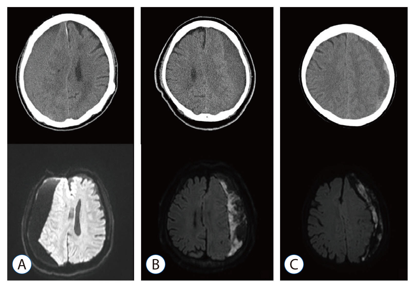
Fig. 1 Appearance of subdural hematomas on diffusion-weighted magnetic resonance imaging (DW-MRI) and preoperative computed tomography (CT). A: Homogeneous density on CT and homogeneous signal intensity on DW-MRI. B: Homogeneous density on CT and mixed signal intensity on DW-MRI. C: Heterogeneous density on CT and mixed signal intensity on DW-MRI.
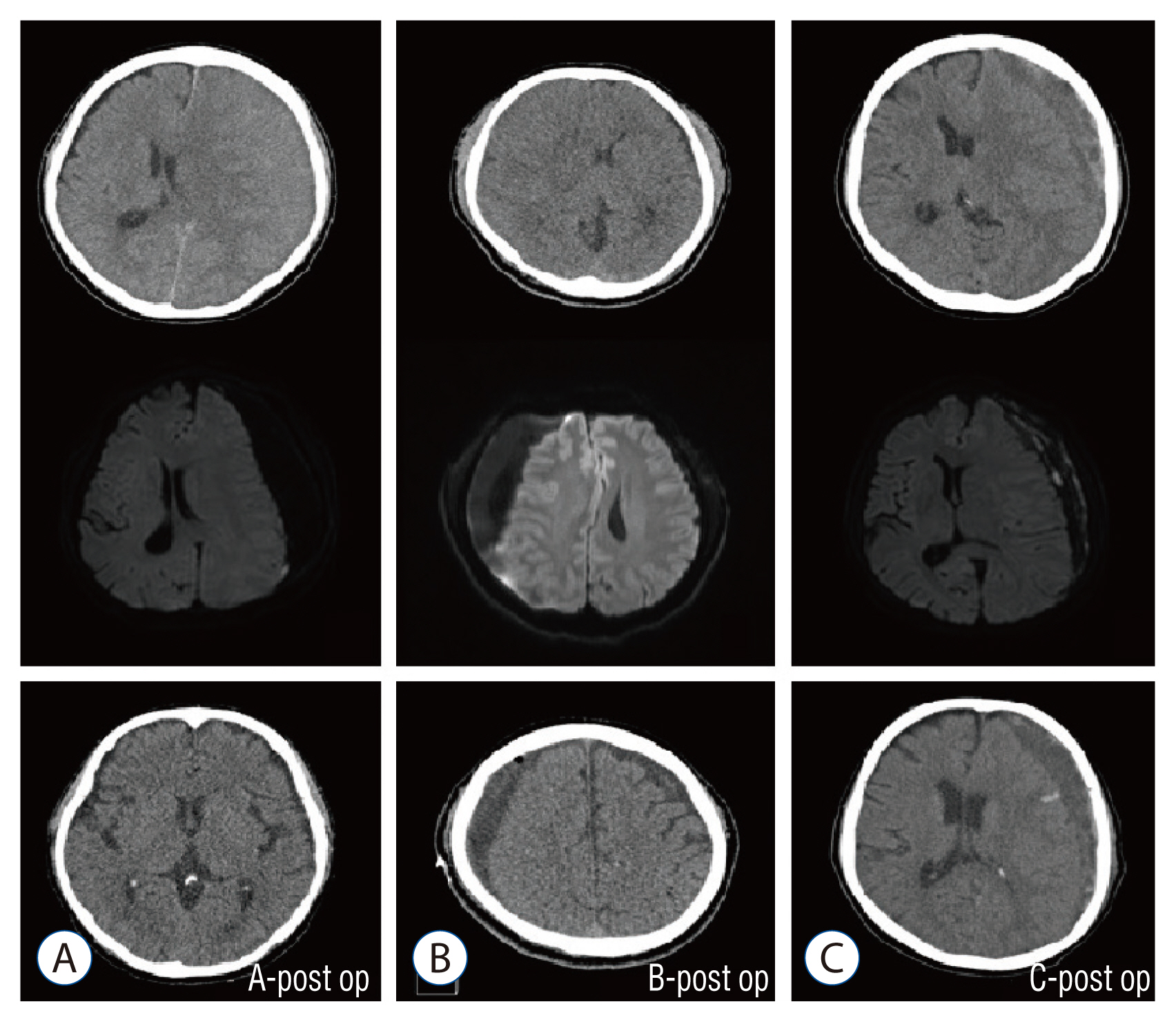
Fig. 2 Postoperative computed tomography of each group in this study. A : Favorable surgical outcomes. B and C : Poor surgical outcomes.
Reference
-
References
1. Amirjamshidi A, Abouzari M, Eftekhar B, Rashidi A, Rezaii J, Esfandiari K, et al. Outcomes and recurrence rates in chronic subdural haematoma. Br J Neurosurg. 21:272–275. 2007.
Article2. Aspegren OP, Åstrand R, Lundgren MI, Romner B. Anticoagulation therapy a risk factor for the development of chronic subdural hematoma. Clin Neurol Neurosurg. 115:981–984. 2013.
Article3. Augustin M, Bammer R, Simbrunner J, Stollberger R, Hartung HP, Fazekas F. Diffusion-weighted imaging of patients with subacute cerebral ischemia: comparison with conventional and contrast-enhanced MR imaging. AJNR Am J Neuroradiol. 21:1596–1602. 2000.4. Borger V, Vatter H, Oszvald Á, Marquardt G, Seifert V, Güresir E. Chronic subdural haematoma in elderly patients: a retrospective analysis of 322 patients between the ages of 65–94 years. Acta Neurochir (Wien). 154:1549–1554. 2012.
Article5. Fainardi E, Borrelli M, Saletti A, Sarubbo S, Roversi G, Bernardoni A, et al. Temporal changes in perihematomal apparent diffusion coefficient values during the transition from acute to subacute phases in patients with spontaneous intracerebral hemorrhage. Neuroradiology. 55:145–156. 2013.
Article6. Goto H, Ishikawa O, Nomura M, Tanaka K, Nomura S, Maeda K. Magnetic resonance imaging findings predict the recurrence of chronic subdural hematoma. Neurol Med Chir (Tokyo). 55:173–178. 2015.
Article7. Hosoda K, Tamaki N, Masumura M, Matsumoto S, Maeda F. Magnetic resonance images of chronic subdural hematomas. J Neurosurg. 67:677–683. 1987.
Article8. Jamjoom A, Nelson R, Stranjalis G, Wood S, Chissell H, Kane N, et al. Outcome following surgical evacuation of traumatic intracranial haematomas in the elderly. Br J Neurosurg. 6:27–32. 1992.
Article9. Ko BS, Lee JK, Seo BR, Moon SJ, Kim JH, Kim SH. Clinical analysis of risk factors related to recurrent chronic subdural hematoma. J Korean Neurosurg Soc. 43:11–15. 2008.
Article10. Kuwahara S, Fukuoka M, Koan Y, Miyake H, Ono Y, Moriki A, et al. Subdural hyperintense band on diffusion-weighted imaging of chronic subdural hematoma indicates bleeding from the outer membrane. Neurol Med Chir (Tokyo). 45:125–131. 2005.
Article11. Kuwahara S, Fukuoka M, Koan Y, Miyake H, Ono Y, Moriki A, et al. Diffusion-weighted imaging of traumatic subdural hematoma in the subacute stage. Neurol Med Chir (Tokyo). 45:464–469. 2005.
Article12. Kuwahara S, Miyake H, Fukuoka M, Koan Y, Ono Y, Moriki A, et al. Diffusion-weighted magnetic resonance imaging of organized subdural hematoma--case report. Neurol Med Chir (Tokyo). 44:376–379. 2004.
Article13. Kwon TH, Park YK, Lim DJ, Cho TH, Chung YG, Chung HS, et al. Chronic subdural hematoma: evaluation of the clinical significance of postoperative drainage volume. J Neurosurg. 93:796–799. 2000.
Article14. Leroy HA, Aboukaïs R, Reyns N, Bourgeois P, Labreuche J, Duhamel A, et al. Predictors of functional outcomes and recurrence of chronic subdural hematomas. J Clin Neurosci. 22:1895–1900. 2015.
Article15. Markwalder TM. Chronic subdural hematomas: a review. J Neurosurg. 54:637–645. 1981.
Article16. Nakaguchi H, Tanishima T, Yoshimasu N. Factors in the natural history of chronic subdural hematomas that influence their postoperative recurrence. J Neurosurg. 95:256–262. 2001.
Article17. Ohba S, Kinoshita Y, Nakagawa T, Murakami H. The risk factors for recurrence of chronic subdural hematoma. Neurosurg Rev. 36:145–149. discussion 149–150. 2013.
Article18. Richter HP, Klein HJ, Schäfer M. Chronic subdural haematomas treated by enlarged burr-hole craniotomy and closed system drainage. Retrospective study of 120 patients. Acta Neurochir (Wien). 71:179–188. 1984.
Article19. Ro HW, Park SK, Jang DK, Yoon WS, Jang KS, Han YM. Preoperative predictive factors for surgical and functional outcomes in chronic subdural hematoma. Acta Neurochir (Wien). 158:135–139. 2016.
Article20. Santarius T, Kirkpatrick PJ, Ganesan D, Chia HL, Jalloh I, Smielewski P, et al. Use of drains versus no drains after burr-hole evacuation of chronic subdural haematoma: a randomised controlled trial. Lancet. 374:1067–1073. 2009.
Article21. Santarius T, Kirkpatrick PJ, Kolias AG, Hutchinson PJ. Working toward rational and evidence-based treatment of chronic subdural hematoma. Clin Neurosurg. 57:112–122. 2010.22. Sipponen JT, Sepponen RE, Sivula A. Chronic subdural hematoma: demonstration by magnetic resonance. Radiology. 150:79–85. 1984.
Article23. Tsutsumi K, Maeda K, Iijima A, Usui M, Okada Y, Kirino T. The relationship of preoperative magnetic resonance imaging findings and closed system drainage in the recurrence of chronic subdural hematoma. J Neurosurg. 87:870–875. 1997.
Article24. Tugcu B, Tanriverdi O, Baydin S, Hergunsel B, Günaldi Ö, Ofluoglu E, et al. Can recurrence of chronic subdural hematoma be predicted? A retrospective analysis of 292 cases. J Neurol Surg A Cent Eur Neurosurg. 75:37–41. 2014.
Article
- Full Text Links
-
- Actions
-
Cited
- CITED
-
- Close
- Share
-
- Similar articles
-
- RE: Diffusion-Weighted Imaging of Prostate Cancer: How Can We Use It Accurately?
- Bone Involvement of Diffuse Large B Cell Lymphoma (DLBCL) Showing Unusual Manifestations Mimicking Chronic Osteomyelitis in a 58-Year-Old Man: Case Report and Clinical Application of Diffusion Weighted Magnetic Resonance Imaging
- Diffusion-Weighted Magnetic Resonance Imaging of Spine
- The Value of PROPELLER Diffusion-Weighted Image in the Detection of Cholesteatoma
- Diffusion-Weighted Magnetic Resonance Imaging Findings in a Patient with Trigeminal Ganglioneuroma
