Application of digital implant system on implant treatment with “all-on-4†concept
- Affiliations
-
- 1Department of Prosthodontics, Wonju College of Medicine, Yonsei University, Wonju, Republic of Korea. smj3@yonsei.ac.kr
- 2Department of Oral and Maxillofacial Surgery, Wonju College of Medicine, Yonsei University, Wonju, Republic of Korea.
- KMID: 2402992
- DOI: http://doi.org/10.4047/jkap.2018.56.1.88
Abstract
- Recently, digital implant systems are expanding its influence in dental area. Due to technical improvement, they jumped over their limits nowadays. We can use these newest systems to treat edentulous patient, from implant surgery to fabrication of prosthesis. In this case, The patient was a fifty years old female. She had a full edentulous ridge on mandible and wanted to reconstruct occlusion with using implants. We planned to use digital implant system with "all-on-4" concept on mandible and produced surgical guide for flapless implant surgery. After the surgery, we tried to fabricate full arch prosthesis just using a digital devices and confirmed satisfying result.
Keyword
MeSH Terms
Figure
-
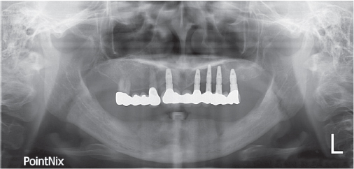
Fig. 1 Pre-operative panoramic radiograph.
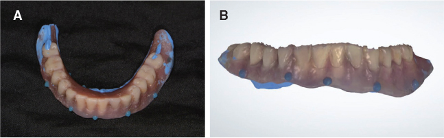
Fig. 2 Scan of existing denture. (A) Radiolucent resin marker on the old denture, (B) 3D scan image.

Fig. 3 3D Image of mandible. (A) Denture biting image, (B) Deletion of denture image, (C) Final mandibular Image.

Fig. 4 Positioning of implants. (A) Merged image, (B) Deletion of bone image.

Fig. 5 Pre-operative preparations. (A) Surgical guide, (B) Temporary abutments and temporary restoration.
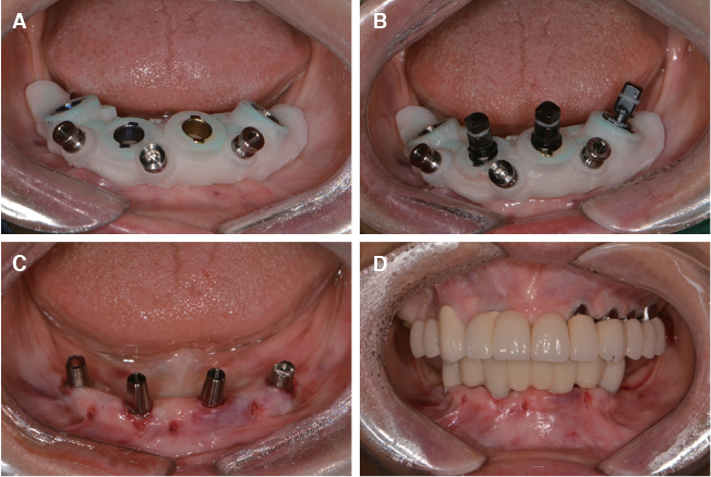
Fig. 6 Flapless implant surgery. (A) Surgical guide, (B) Positioning of the implant, (C) Connection of the abutments, (D) Cementation of the temporary restoration.
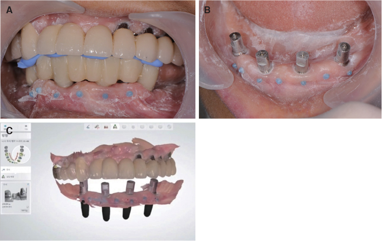
Fig. 7 3D image for final prosthesis. (A) Resin marker on gingiva, (B) Scan body, (C) Final image.
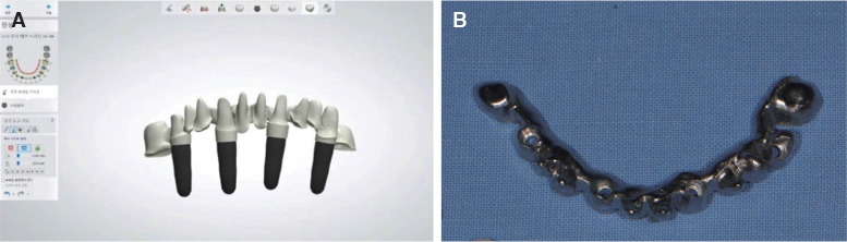
Fig. 8 Fabrication of framework. (A) 3D image, (B) Milled metal framework.
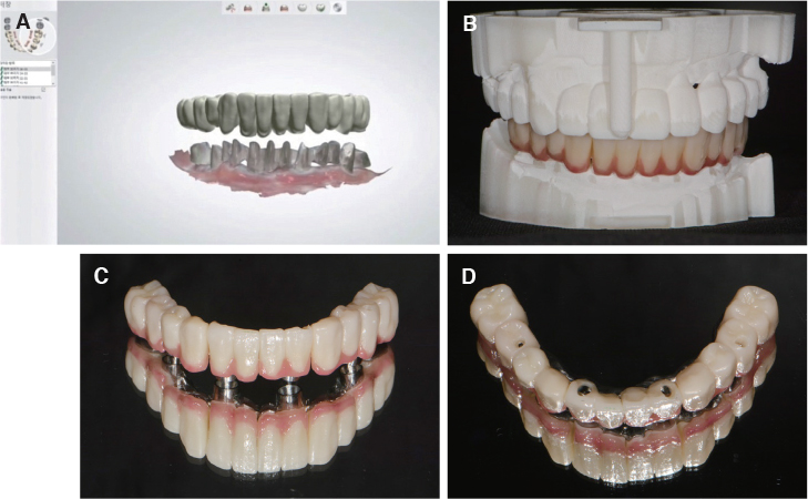
Fig. 9 Final prosthesis. (A) Zirconia shell image, (B) Resin model, (C) Frontal view, (D) Occlusal view.
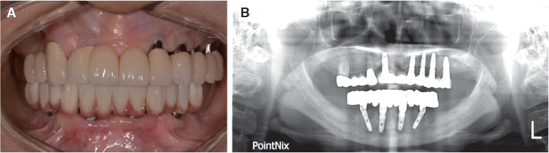
Fig. 10 After treatment. (A) Intraoral photo, (B) Panoramic radiograph.
Reference
-
1. Atwood DA. Postextraction changes in the adult mandible as illustrated by microradiographs of midsagittal sections and serial cephalometric roentgenograms. J Prosthet Dent. 1963; 13:810–824.
Article2. Wical KE, Swoope CC. Studies of residual ridge resorption. I. Use of panoramic radiographs for evaluation and classification of mandibular resorption. J Prosthet Dent. 1974; 32:7–12.
Article3. Cawood JI, Howell RA. A classification of the edentulous jaws. Int J Oral Maxillofac Surg. 1988; 17:232–236.
Article4. Zarb GA, Bolender CL, Ecker S, Jacob R, Fenton A, Mericske-Stern R. Prosthodontic treatment for edentulous patients. 12th ed. St. Louis: Elsevier Mosby;2004.5. Wismeijer D, van Waas MA, Kalk W. Factors to consider in selecting an occlusal concept for patients with implants in the edentulous mandible. J Prosthet Dent. 1995; 74:380–384.
Article6. Brånemark PI, Svensson B, van Steenberghe D. Ten-year survival rates of fixed prostheses on four or six implants ad modum Brånemark in full edentulism. Clin Oral Implants Res. 1995; 6:227–231.
Article7. Kopp CD. Brånemark osseointegration. Prognosis and treatment rationale. Dent Clin North Am. 1989; 33:701–731.8. Krekmanov L, Kahn M, Rangert B, Lindström H. Tilting of posterior mandibular and maxillary implants for improved prosthesis support. Int J Oral Maxillofac Implants. 2000; 15:405–414.9. Kim KS, Kim YL, Bae JM, Cho HW. Biomechanical comparison of axial and tilted implants for mandibular full-arch fixed prostheses. Int J Oral Maxillofac Implants. 2011; 26:976–984.10. Choi BH, Jeong SM. Digital flapless implantology. Seoul, Korea: Jisung Pub.;2015.11. Maló P, de Araújo Nobre M, Lopes A, Francischone C, Rigolizzo M. “All-on-4” immediate-function concept for completely edentulous maxillae: a clinical report on the medium (3 years) and long-term (5 years) outcomes. Clin Implant Dent Relat Res. 2012; 14:e139–e150.
Article12. Katsoulis J, Müller P, Mericske-Stern R, Blatz MB. CAD/CAM fabrication accuracy of long- vs. short-span implantsupported FDPs. Clin Oral Implants Res. 2015; 26:245–249.
Article13. Alghazzawi TF. Advancements in CAD/CAM technology: Options for practical implementation. J Prosthodont Res. 2016; 60:72–84.
Article
- Full Text Links
-
- Actions
-
Cited
- CITED
-
- Close
- Share
-
- Similar articles
-
- Mini-implant with additional retentive structure by using digital method
- A Completely Cast-Free, Digital Workflow for Fabricating an Implant-Supported Prosthesis with a New Coded Healing Abutment
- All-on-6 implant fixed prosthesis restoration with fulldigital system on edentulous patient: A case report
- Full mouth rehabilitation of an edentulous patient using maxillary complete denture and mandibular implant supported fixed prostheses: a case report
- Digital intraoral impression for immediate provisional restoration of maxillary single implant: A case report
