J Korean Soc Spine Surg.
2017 Sep;24(3):162-168. 10.4184/jkss.2017.24.3.162.
Relationship Between Low Back Pain and the Size and Density of the Erector Spinae Muscle and Multifidus Muscle Using CT Imaging in a Selected Community-Based Population
- Affiliations
-
- 1Department of Orthopaedic Surgery, Daegu Catholic University Medical Center, Daegu, Korea. bong@cu.ac.kr
- KMID: 2402829
- DOI: http://doi.org/10.4184/jkss.2017.24.3.162
Abstract
- STUDY DESIGN: Case-control study (retrospective comparative study).
OBJECTIVES
The purpose of this study was to define the relationship between low back pain (LBP) and the cross-sectional area (CSA) and density of the erector spinae muscle (ESM) and isolated multifidus muscle (IMM) on computed tomography (CT) scans of patients with a chief complaint other than LBP. SUMMARY OF LITERATURE REVIEW: Most previous studies have focused on radiographic data from patients with a chief complaint of LBP, rather than on radiographic data from patients with a chief complaint other than LBP.
MATERIALS AND METHODS
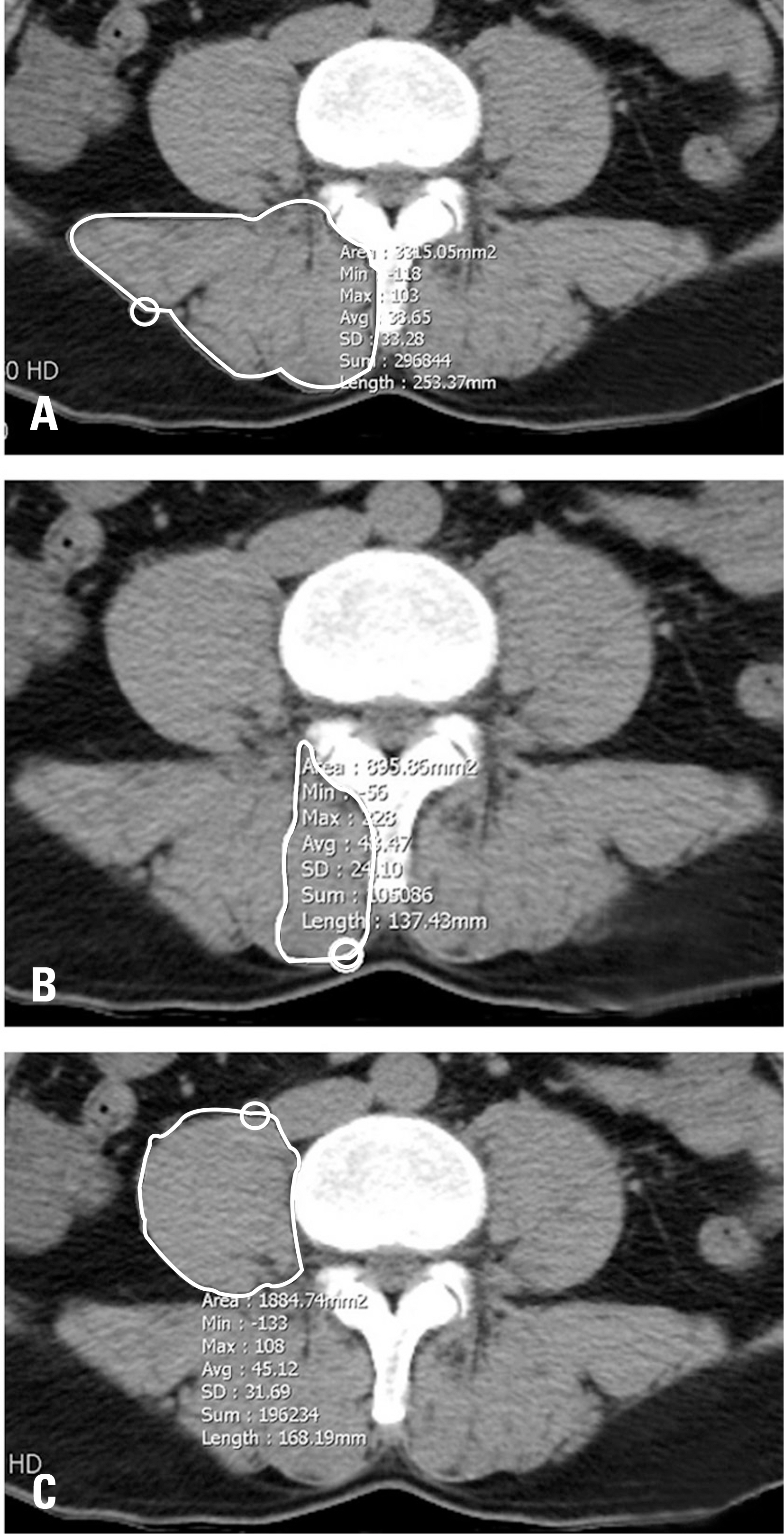
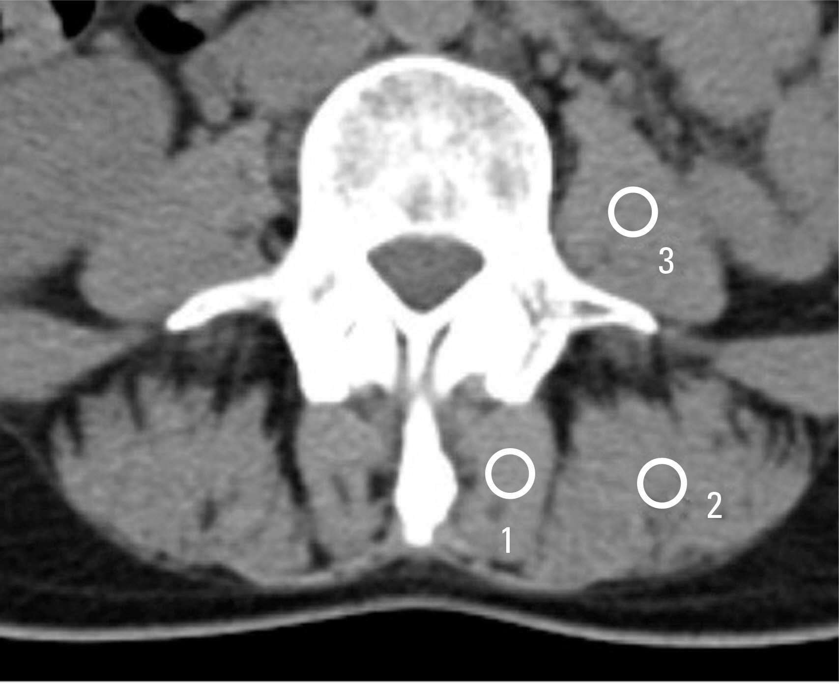
This retrospective study included 475 patients who underwent CT scans between January 1, 2010 and December 31, 2010. The CSA and density of the ESM, IMM, and the psoas muscle (PM) were obtained. All measurements were calculated as the ratio of each muscle. The relationships between the CSA of each muscle and both types of LBP were analyzed.
RESULTS
The ESM-to-PM ratio in terms of density was 1.227±0.797 in the LBP group and 0.645±0.732 in the non-LBP group (p=0.174). The IMM-to-PM ratio in terms of density was 0.664±0.515 in the LBP group and 0.806±0.518 in the non-LBP group (p=0.007).
CONCLUSIONS
The IMM was more relevant to LBP than the ESM of the back, and density was more relevant to LBP than the CSA of regular muscles. The IMM was more useful than the ESM for analyzing LBP.
MeSH Terms
Figure
Reference
-
1. Mayer TG, Vanharanta H, Gatchel RJ, et al. Comparison of CT scan muscle measurements and isokinetic trunk strength in postoperative patients. Spine (Phila Pa 1976). 1989; 14:33–6.
Article2. Kamaz M, Kireşi D, Oğuz H, et al. CT measurement of trunk muscle areas in patients with chronic low back pain. Diagn Interv Radiol. 2007; 13:144–8.3. Storheim K, Holm I, Gunderson R, et al. The effect of comprehensive group training on cross-sectional area, density, and strength of paraspinal muscles in patients sick-listed for subacute low back pain. J Spinal Disord Tech. 2003; 16:271–9.
Article4. Hodges PW. The role of the motor system in spinal pain: implications for rehabilitation of the athlete following lower back pain. J Sci Med Sport. 2000; 3:243–53.
Article5. Sato H, Kikuchi S. The natural history of radiographic instability of the lumbar spine. Spine (Phila Pa 1976). 1993; 18:2075–9.
Article6. Alam WCA. Radiological evaluation of lumbar intervertebral instability. Ind J Aerospace Med. 2002; 46:48–53.7. Kjaer P, Bendix T, Sorensen JS, et al. Are MRI-defined fat infiltrations in the multifidus muscles associated with low back pain? BMC Medicine. 2007; 5:2.
Article8. Mattila M, Hurme M, Alaranta H, et al. The multifidus muscle in patients with lumbar disc herniation: a histochemical and morphometric analysis of intraoperative biopsies. Spine. 1986; 11:732–8.9. Sihvonen T, Herno A, Paljärvi L, et al. Local denervation atrophy of paraspinal muscles in postoperative failed back syndrome. Spine (Phila Pa 1976). 1993; 18:575–81.
Article10. Kay AG. An extensive literature review of the lumbar multifidus: anatomy. J Manual Manipulative Ther. 2000; 8:102–14.
Article11. Kuorinka I, Jonsson B, Kilbom A, et al. Standardized Nordic questionnaires for the analysis of musculoskeletal symptoms. Appl Ergon. 1987; 18:233–7.12. Han JS, Ahn JY, Goel VK, et al. CT-based geometric data of human spine musculature, Part I. Japanese patients with chronic low back pain. J Spinal Disord Tech. 1992; 5:448–58.
Article13. Hides JA, Stokes MJ, Saide M, et al. Evidence of lumbar multifidus muscle wasting ipsilateral to symptoms in patients with acute/subacute low back pain. Spine (Phila Pa 1976). 1994; 19:165–72.
Article14. Käser L, Mannion AF, Rhyner A, et al. Active therapy for chronic low back pain: part 2. Effects on paraspinal muscle cross-sectional area, fiber type size, and distribution. Spine (Phila Pa 1976). 2001; 26:909–19.15. Akima H, Kuno S, Takahashi H, et al. The use of magnetic resonance images to investigate the influence of recruitment on the relationship between torque and cross-sectional area in human muscle. Eur J Apply Physiol. 2000; 83:475–80.
Article16. Keller A, Gunderson R, Reikerås O, et al. Reliability of computed tomography measurements of paraspinal muscle cross-sectional area and density in patients with low back pain. Spine (Phila Pa 1976). 2003; 28:1455–60.17. Cholewicki J, McGill SM. Mechanical stability of the in vivo lumbar spine: implications for injury and chronic low back pain. Clin Biomech. 1996; 11:1–15.
Article18. Crisco JJ 3rd, Panjabi MM. The intersegmental and multisegmental muscles of the lumbar spine. A biomechanical model comparing lateral stabilizing potential. Spine (Phila Pa 1976). 1991; 16:793–9.19. Le Huec JC, Mathews H, Basso Y, et al. Clinical results of Maverick lumbar total disc replacement: two-year prospective follow-up. Orthop Clin North Am. 2005; 36:315–22.
Article20. Panjabi MM. The stabilizing system of the spine. Part I. Function, dysfunction, adaptation, and enhancement. J Spinal Disord Tech. 1992; 5:383–9.
Article21. Mannion AF, Käser L, Weber E, et al. Influence of age and duration of symptoms on fibre type distribution and size of the back muscles in chronic low back patients. Eur Spine J. 2000; 9:273–81.22. Danneels LA, Vanderstraeten GG, Cambier DC, et al. CT imaging of trunk muscles in chronic low back pain patients and healthy control subjects. Eur Spine J. 2000; 9:266–72.
Article23. Parkkola R, Rytökoski U, Kormano M. Magnetic resonance imaging of the discs and trunk muscles in patients with chronic low back pain and healthy control subjects. Spine (Phila Pa 1976). 1993; 18:830–6.
Article24. Flicker PL, Fleckenstein JL, Ferry K, et al. Lumbar muscle usage in chronic low back pain. Magnetic resonance image evaluation. Spine (Phila Pa 1976). 1993; 18:582–6.25. Kader DF, Wardlaw D, Smith FW. Correlation between the MRI changes in the lumbar multifidus muscles and leg pain. Clin Radiol. 2000; 55:145–9.
Article26. Santaguida PL, McGill SM. The psoas major muscle: a three-dimensional geometric study. J Biomech. 1995; 28:339–45.
Article27. McGill S, Juker D, Kropf P. Quantitative intramuscular myoelectric activity of quadratus lumborum during a wide variety of tasks. Clin Biomech. 1996; 11:170–2.
Article28. Pietrek M, Sheikhzadeh A, Nordin M, et al. Biomechanical modeling of intra-abdominal pressure generation should include the transversus abdominis. J Biomech. 2000; 33:787–90.29. Hides JA, Richardson CA, Jull GA. Magnetic resonance imaging and ultrasonography of the lumbar multifidus muscle: comparison of two different modalities. Spine (Phila Pa 1976). 1995; 20:54–8.
- Full Text Links
- Actions
-
Cited
- CITED
-
- Close
- Share
- Similar articles
-
- Facilitating Effects of Fast and Slope Walking on Paraspinal Muscles
- Association between Cross-sectional Areas of Lumbar Muscles on Magnetic Resonance Imaging and Chronicity of Low Back Pain
- Relationship between Lumbar Disc Degeneration and Back Muscle Degeneration
- The Variation in the Lumbar Facet Joint Orientation in an Adult Asian Population and Its Relationship with the Cross-Sectional Area of the Multifidus and Erector Spinae
- Sarcopenia in elderly patients with chronic low back pain


