Korean J Ophthalmol.
2018 Feb;32(1):38-44. 10.3341/kjo.2017.0031.
Factors Affecting ISNT Rule Satisfaction in Normal and Glaucomatous Eyes
- Affiliations
-
- 1Department of Ophthalmology, Seoul National University College of Medicine, Seoul, Korea. xcski@hanmail.net
- 2Department of Ophthalmology, Seoul National University Hospital, Seoul, Korea.
- 3Department of Ophthalmology, SMG-SNU Boramae Medical Center, Seoul, Korea.
- KMID: 2402718
- DOI: http://doi.org/10.3341/kjo.2017.0031
Abstract
- PURPOSE
To determine the factors that influence the satisfaction of the "˜ISNT rule' (neural rim width: inferior ≥ superior ≥ nasal ≥ temporal) in normal and glaucomatous eyes.
METHODS
The medical records of patients that visited Boramae Medical Center, Seoul, Korea, were reviewed. Each group of normal and glaucomatous eyes was divided into subgroups based on whether or not they satisfied the ISNT rule. ISNT rule assessment was performed by measuring the rim width with stereoscopic optic disc photographs using ImageJ software. Logistic regression analysis was performed to determine the factors that affect ISNT rule satisfaction.
RESULTS
Seventy-seven normal eyes and 97 glaucomatous eyes were included in the study. The ISNT rule was intact in 59 (76.6%) of the normal eyes and was violated in 71 (73.2%) of the glaucomatous eyes. Logistic regression analysis revealed a significant influence of axial length in violation of the ISNT rule in the normal eye group, while the mean deviation value was a significant factor for violation of the ISNT rule in the glaucomatous eye group.
CONCLUSIONS
The ISNT rule should be cautiously applied when evaluating normal eyes with long axial lengths. In addition, the ISNT rule might not be as effective for detecting early glaucoma.
Keyword
MeSH Terms
Figure
-
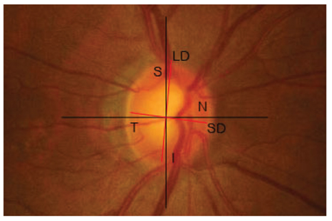
Fig. 1 Clinical assessment of the normal optic disc. Optic disc center was defined as the intersection of the longest diameter (LD) and shortest diameter (SD) of the optic disc. Superior (S) and inferior (I) rims were defined as the vertical line that passes through the optic disc center. Nasal (N) and temporal (T) rims were defined as the horizontal line that passes through the optic disc center.
Reference
-
1. Quigley HA, Katz J, Derick RJ, et al. An evaluation of optic disc and nerve fiber layer examinations in monitoring progression of early glaucoma damage. Ophthalmology. 1992; 99:19–28.
Article2. Sommer A, Quigley HA, Robin AL, et al. Evaluation of nerve f iber layer assessment. Arch Ophthalmol. 1984; 102:1766–1771.3. Jonas JB, Fernandez MC, Sturmer J. Pattern of glaucomatous neuroretinal rim loss. Ophthalmology. 1993; 100:63–68.
Article4. Tuulonen A, Airaksinen PJ. Initial glaucomatous optic disk and retinal nerve fiber layer abnormalities and their progression. Am J Ophthalmol. 1991; 111:485–490.
Article5. Chan EW, Liao J, Wong R, et al. Diagnostic performance of the ISNT rule for glaucoma based on the Heidelberg Retinal Tomograph. Transl Vis Sci Technol. 2013; 2:2.
Article6. Lisboa R, Leite MT, Zangwill LM, et al. Diagnosing preperimetric glaucoma with spectral domain optical coherence tomography. Ophthalmology. 2012; 119:2261–2269.
Article7. Nayak NV, Berezina TL, Fechtner RD, et al. Effect of age and disc size on rim order rules by Heidelberg Retina Tomograph. J Glaucoma. 2015; 24:377–382.
Article8. Arnalich-Montiel F, Munoz-Negrete FJ, Rebolleda G, et al. Cup-to-disc ratio: agreement between slit-lamp indirect ophthalmoscopic estimation and stratus optical coherence tomography measurement. Eye (Lond). 2007; 21:1041–1049.
Article9. Carpel EF, Engstrom PF. The normal cup-disk ratio. Am J Ophthalmol. 1981; 91:588–597.
Article10. Jonas JB, Gusek GC, Naumann GO. Optic disc, cup and neuroretinal rim size, configuration and correlations in normal eyes. Invest Ophthalmol Vis Sci. 1988; 29:1151–1158.11. Harizman N, Oliveira C, Chiang A, et al. The ISNT rule and differentiation of normal from glaucomatous eyes. Arch Ophthalmol. 2006; 124:1579–1583.
Article12. Wang Y, Xu L, Jonas JB. Shape of the neuroretinal rim and its correlations with ocular and general parameters in adult chinese: the beijing eye study. Am J Ophthalmol. 2007; 144:462–464.
Article13. Sihota R, Srinivasan G, Dada T, et al. Is the ISNT rule violated in early primary open-angle glaucoma: a scanning laser tomography study. Eye (Lond). 2008; 22:819–824.14. Morgan JE, Bourtsoukli I, Rajkumar KN, et al. The accuracy of the inferior>superior>nasal>temporal neuroretinal rim area rule for diagnosing glaucomatous optic disc damage. Ophthalmology. 2012; 119:723–730.15. Law SK, Kornmann HL, Nilforushan N, et al. Evaluation of the "IS" rule to differentiate glaucomatous eyes from normal. J Glaucoma. 2016; 25:27–32.
Article16. Jonas JB, Gusek GC, Naumann GO. Optic disc morphometry in chronic primary open-angle glaucoma. I. Morphometric intrapapillary characteristics. Graefes Arch Clin Exp Ophthalmol. 1988; 226:522–530.17. Jonas JB, Dichtl A. Optic disc morphology in myopic primary open-angle glaucoma. Graefes Arch Clin Exp Ophthalmol. 1997; 235:627–633.
Article18. Jonas JB, Gusek GC, Naumann GO. Optic disk morphometry in high myopia. Graefes Arch Clin Exp Ophthalmol. 1988; 226:587–590.
Article19. Park HY, Lee K, Park CK. Optic disc torsion direction predicts the location of glaucomatous damage in normal-tension glaucoma patients with myopia. Ophthalmology. 2012; 119:1844–1851.
Article
- Full Text Links
-
- Actions
-
Cited
- CITED
-
- Close
- Share
-
- Similar articles
-
- Discriminating Between Normal and Glaucomatous Eyes Using the Modified ISNT Rule
- Applicability of ISNT Rule Using Bruch's Membrane Opening-based Optic Nerve Head Parameters
- Peripapillary Atrophy in Asymmetric Glaucoma
- Diverse Types of Glaucoma in Patients with Pseudoexfoliation Syndrome: Normal Pressure Glaucoma
- Comparison of Glaucomatous Parameters in Normal, Ocular Hypertensive and Glaucomatous Eyes Using Optical Coherence Tomography 3000
