Anatomical Reduction with Brick-Work Technique in Comminuted Intraarticular Distal Radius Fractures
- Affiliations
-
- 1Department of Orthopedic Surgery, Good Samsun Hospital, Busan, Korea. hc5771@naver.com
- KMID: 2402708
- DOI: http://doi.org/10.12671/jkfs.2018.31.1.1
Abstract
- PURPOSE
This study examined the clinical outcomes of comminuted intraarticular distal radius fractures treated by an anatomical reduction using a brick-work technique.
MATERIALS AND METHODS
Seventeen patients with AO/OTA type 23-C3 distal radius fractures were enrolled in this study. An anatomical reduction of the articular surface was achieved using a brick-work technique through the dorsal approach and dorsal plates were used for fixation. The postoperative functional results were assessed with the range of motion of the wrist and the modified Mayo wrist score (MMWS). In addition, the radial length, radial inclination, volar tilt, and Lidstrom score were evaluated from the radiology results. The mean postoperative follow-up period was 13.6 months.
RESULTS
All patients showed bony union and the mean range of motion of the injured wrists was 94% (92% to 95%) of the uninjured side. The mean MMWS was 85.3, and the functional results were excellent in 12 patients, good in 4, and fair in one at the final follow-up. Based on the final radiographic measurements, the radial length, volar tilt, and radial inclination were 11.4 mm (10.0 to 13.5 mm), 6.6° (−1.8° to 9.2°), and 21.3° (20.1° to 25.7°), respectively. The radiologic results according to the Lidstrom score were excellent in 14 patients and good in three.
CONCLUSION
An anatomical reduction with the brick-work technique is relatively easy, results in a reproducible clinical outcome, and could be a safe and effective treatment option for severe comminuted intraarticular distal radius fractures that are not amenable to volar plate fixation.
Figure
-
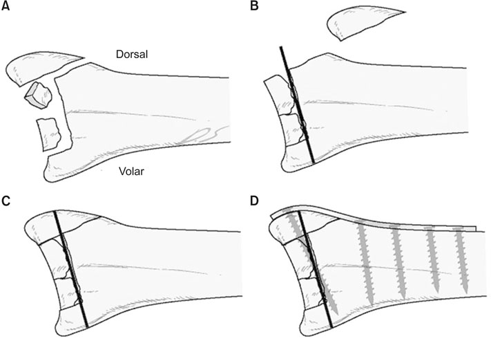
Fig. 1 Schematic diagram of the dorsal plating with brick-work technique. (A) Displaced intraarticular fracture with multi-fragmentary central and dorsal comminution. (B) After elevating the dorsal comminuted fragments, the articular surface was reduced from the volar to dorsal sequence and provisional fixation with a temporary K-wire. (C) Dorsal comminuted fragments were reduced in its original position. (D) Final application of dorsal contoured plate and screws.
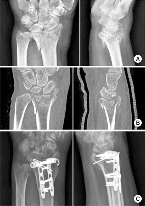
Fig. 2 Case of a 74-year-old male with an intraarticular distal radius fracture. Posteroanterior and lateral radiographs (A), and computed tomography images (B) show central and dorsal multi-fragmentary comminution corresponding to an AO/OTA 23-C3 fracture pattern. (C) Postoperative follow-up radiographs after 6 months show complete union.
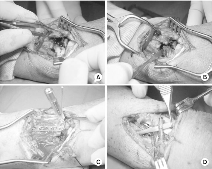
Fig. 3 Intraoperative images of dorsal plating with brick-work technique reduction. (A) The dorsal fragment was elevated for the reduction of displaced volar articular fragments. (B) The articular surface was reduced using periosteal elevator. (C) The plate was fixed using a locking screw. (D) Extensor retinacular sling for the protection of the extensor pollicis longus tendon.
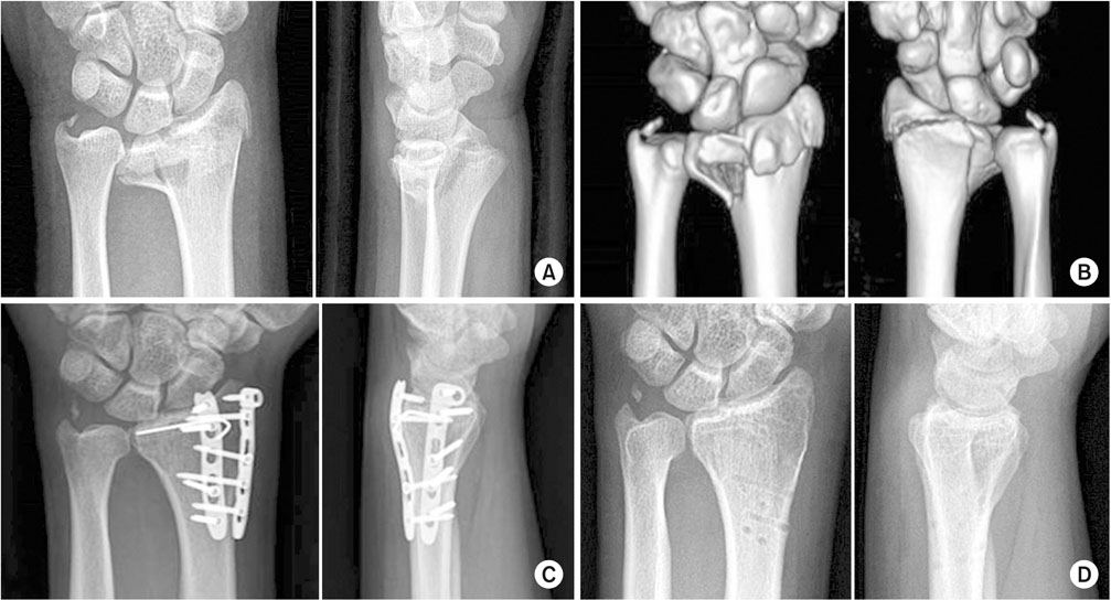
Fig. 4 Case of a 31-year-old male with an intraarticular distal radius fracture with dorsal comminution. Posteroanterior and lateral radiographs (A), and three dimensional reconstructed computed tomography images (B) show dorsally displaced comminuted fragments. (C) Postoperative followup radiographs after 5 months show complete union and maintained radiographic parameters. (D) Final follow-up radiographs after removing the implants at 11 months after surgery.
Reference
-
1. Lidstrom A. Fractures of the distal end of the radius. A clinical and statistical study of end results. Acta Orthop Scand Suppl. 1959; 41:1–118.2. Leung F, Tu YK, Chew WY, Chow SP. Comparison of external and percutaneous pin fixation with plate fixation for intra-articular distal radial fractures. A randomized study. J Bone Joint Surg Am. 2008; 90:16–22.
Article3. Ring D, Jupiter JB, Brennwald J, Büchler U, Hastings H 2nd. Prospective multicenter trial of a plate for dorsal fixation of distal radius fractures. J Hand Surg Am. 1997; 22:777–784.
Article4. Arora R, Lutz M, Hennerbichler A, Krappinger D, Espen D, Gabl M. Complications following internal fixation of unstable distal radius fracture with a palmar locking-plate. J Orthop Trauma. 2007; 21:316–322.
Article5. Rozental TD, Blazar PE. Functional outcome and complications after volar plating for dorsally displaced, unstable fractures of the distal radius. J Hand Surg Am. 2006; 31:359–365.
Article6. Tavakolian JD, Jupiter JB. Dorsal plating for distal radius fractures. Hand Clin. 2005; 21:341–346.
Article7. Song SW. Dorsal plating for distal radius fracture. J Korean Fract Soc. 2008; 21:334–340.
Article8. Ruch DS, Papadonikolakis A. Volar versus dorsal plating in the management of intra-articular distal radius fractures. J Hand Surg Am. 2006; 31:9–16.
Article9. Cooney WP, Bussey R, Dobyns JH, Linscheid RL. Difficult wrist fractures. Perilunate fracture-dislocations of the wrist. Clin Orthop Relat Res. 1987; (214):136–147.10. Kapoor H, Agarwal A, Dhaon BK. Displaced intra-articular fractures of distal radius: a comparative evaluation of results following closed reduction, external fixation and open reduction with internal fixation. Injury. 2000; 31:75–79.
Article11. Wright TW, Horodyski M, Smith DW. Functional outcome of unstable distal radius fractures: ORIF with a volar fixed-angle tine plate versus external fixation. J Hand Surg Am. 2005; 30:289–299.
Article12. Lattmann T, Meier C, Dietrich M, Forberger J, Platz A. Results of volar locking plate osteosynthesis for distal radial fractures. J Trauma. 2011; 70:1510–1518.
Article13. Campbell DA. Open reduction and internal fixation of intra articular and unstable fractures of the distal radius using the AO distal radius plate. J Hand Surg Br. 2000; 25:528–534.
Article14. Lutsky K, McKeon K, Goldfarb C, Boyer M. Dorsal fixation of intra-articular distal radius fractures using 2.4-mm locking plates. Tech Hand Up Extrem Surg. 2009; 13:187–196.
Article15. Hastings H 2nd, Leibovic SJ. Indications and techniques of open reduction. Internal fixation of distal radius fractures. Orthop Clin North Am. 1993; 24:309–326.16. Leibovic SJ, Geissler WB. Treatment of complex intra-articular distal radius fractures. Orthop Clin North Am. 1994; 25:685–706.
Article17. Rozental TD, Beredjiklian PK, Bozentka DJ. Functional outcome and complications following two types of dorsal plating for unstable fractures of the distal part of the radius. J Bone Joint Surg Am. 2003; 85-A:1956–1960.
Article18. Dario P, Matteo G, Carolina C, et al. Is it really necessary to restore radial anatomic parameters after distal radius fractures? Injury. 2014; 45:Suppl 6. S21–S26.
Article19. Yu YR, Makhni MC, Tabrizi S, Rozental TD, Mundanthanam G, Day CS. Complications of low-profile dorsal versus volar locking plates in the distal radius: a comparative study. J Hand Surg Am. 2011; 36:1135–1141.
Article
- Full Text Links
-
- Actions
-
Cited
- CITED
-
- Close
- Share
-
- Similar articles
-
- Distal Radius Fracture treated with Distraction Plate Technique
- Failure of Distal Locking Screws in an Intraarticular Distal Radius Fracture Treated with Volar Locking Plate Fixation
- Treatment of Comminuted - Intraarticular Fracture of Distal Radius using External Fixator: Comparative Study of Treatment by External Fixation with or Without bone Graft
- Treatment of Unstable Intraarticular Fracture of Distal Radius with External Fixator and Minimal open Reduction
- Percutaneous pinning of intraarticular comminuted fracture of the distal radius
