Smartphone Fundoscopy to Detect Retinopathy of Prematurity
- Affiliations
-
- 1Department of Ophthalmology, Dong-A University College of Medicine, Busan, Korea. yhkwon@dau.ac.kr
- KMID: 2401675
- DOI: http://doi.org/10.3341/jkos.2018.59.1.31
Abstract
- PURPOSE
To evaluate the usefulness of fundus images captured with a smartphone camera in retinopathy of prematurity (ROP).
METHODS
We took fundoscopic photographs of 13 premature infants (26 eyes) using a smartphone (an I-phone 5) camera fitted with a 30 D lens (Volk Optical Inc., Mentor, OH, USA) from March 2014 to September 2017 in the neonatal intensive care units of Dong-A University Hospital. A hand-held smartphone camera with a 30 D lens was used to record the fundus in video mode. Fundus photographs were then captured from the video film.
RESULTS
Four premature infants were diagnosed with ROP and were successfully photographed via smartphone fundoscopy. The photographs showed the optic disc, retinal arteries and veins, and the posterior pole of the retina. The photographs were simply saved as image files and uploaded to our electronic medical record system.
CONCLUSIONS
Smartphone fundoscopy to document ROP does not capture the peripheral retina but can be useful in hospitals lacking expensive imaging equipment. The technique may be useful in terms of documentation, education, and telemedicine.
MeSH Terms
Figure
-
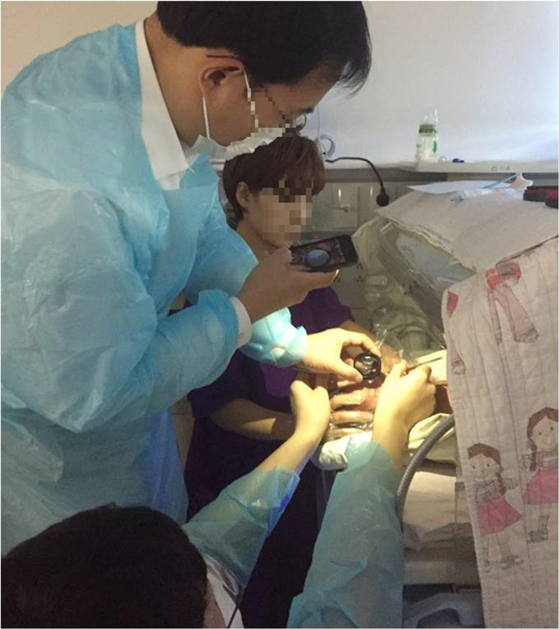
Figure 1. Smartphone fundoscopy technique to detect retinopathy of prematurity. A hand-held smartphone and a 30 D lens were used to take the fundus photograph.

Figure 2. Smartphone fundus photographs of case 1 (29 weeks 6 days, 1,414 g birth weight), the right eye (A-F) and the left eye (G-L). (B, G) Posterior pole of both eyes, and (A, H) posterior zone II area of retina before laser treatment. (C, D, I, J) Posterior pole and posterior zone II area at 2 weeks after treatment. (E, F, K, L) 10 weeks after treatment. Photos show regression of extra-retinal fibrovascular proliferation (E, L) and laser scars on the left eye (L). In case of the right eye, we couldn't take photo showing regression of extraretinal fibrovascular proliferation and laser scars (E).
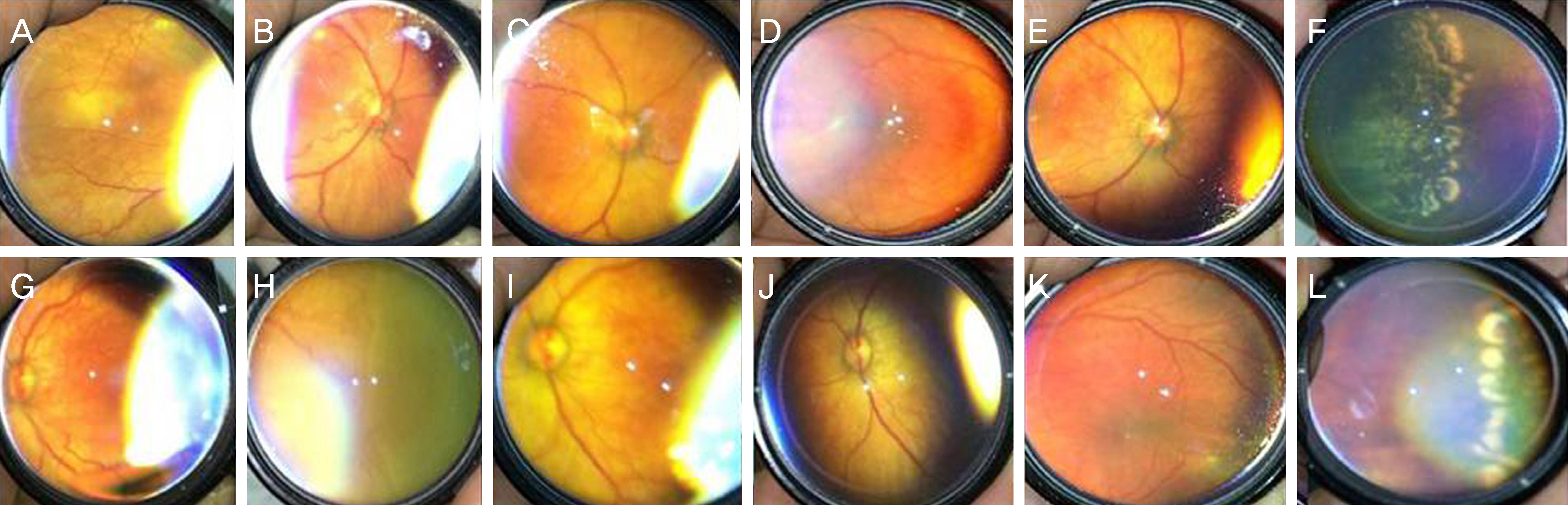
Figure 3. Smartphone fundus photographs of case 4 (26 weeks 5 days, 1,030 g birth weight), the right eye (A-F), and the left eye (G-L). (B, G) Posterior pole of prematurity, and (A, H) posterior zone II area of the both eyes before laser treatment. (C, I) Posterior pole of prematurity at 6 weeks after treatment. Photos of 4 months after treatment (D, E, J, K) show improvement of plus sign. Photos of 6 months after treatment (F, L) show laser scars.
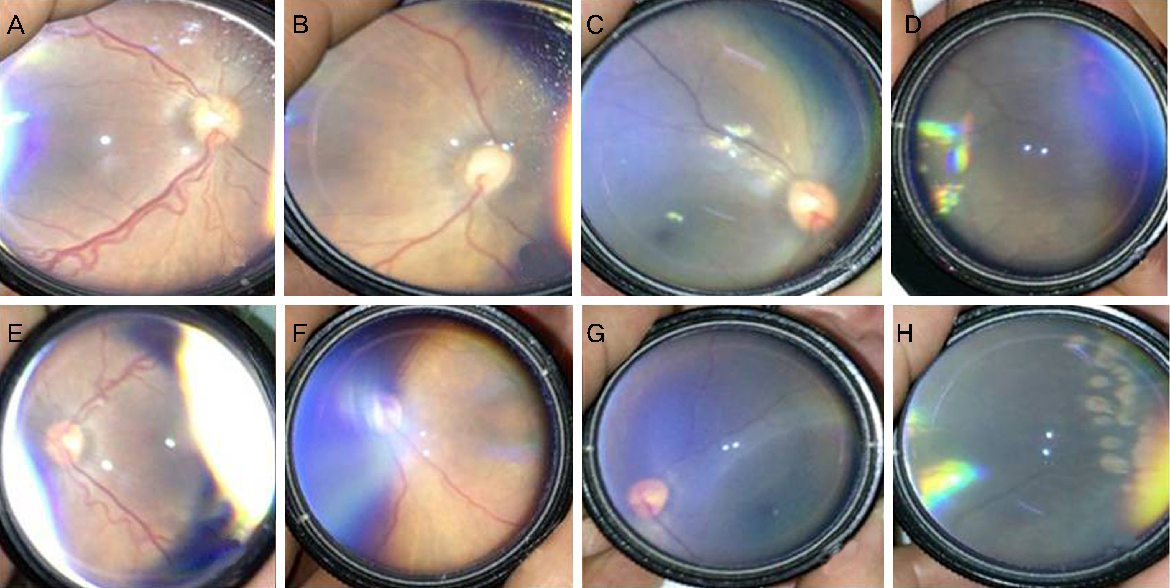
Figure 4. Smartphone fundus photographs of case 7 (27 weeks 6 days, 1,002 g birth weight), the right eye (A-D), and the left eye (E-H). (A, E) Posterior pole of the both eyes before laser treatment. (B, F) Posterior pole at 1 week after treatment. (C, D, G, H) Posterior pole and posterior zone II area at 10 weeks after treatment. (H) Laser scars on the left eye at 10 weeks after laser treatment. In case of the right eye, we couldn't take photo showing laser scars (D).

Figure 5. Smartphone fundus photographs of case 12 (28 weeks, 790 g birth weight), the right eye (A-D), and the left eye (E-H). (B, E) Posterior pole and (A, F) posterior zone II area and ridge. (C, G) Posterior pole 4 days after treatment, and show decreased vessel tortuosity. (D, H) Posterior pole at 1 week after treatment. Treatment after 4 days and 1 week, we couldn't take photo showing regression of retinopathy of prematurity stage and laser scars (C, D, G, H).
Reference
-
References
1. Lord RK, Shah VA, San Filippo AN, Krishna R. Novel uses of smartphones in ophthalmology. Ophthalmol. 2010; 117:1274.
Article2. Zvornicanin E, Zvornicanin J, Hadziefendic B. The use of smart phones in ophthalmol. Acta Inform Med. 2014; 22:206–9.3. Good WV; Early Treatment for Retinopathy of Prematurity Cooperative Group. Final results of the Early Treatment for Retinopathy of Prematurity (ETROP) randomized trial. Trans Am Ophthalmol Soc. 2004; 102:233–48. discussion 248–50.4. Ells AL, Holmes JM, Astle WF, et al. Telemedicine approach to screening for severe retinopathy of prematurity: a pilot study. Ophthalmol. 2003; 110:2113–7.5. Oluleye TS, Rotimi-Samuel A, Adenekan A. Mobile phone for abdominal of prematurity screening in Lagos, Nigeria, sub-Saharan Africa. Eur J Ophthalmol. 2016; 26:92–4.6. Yoon HS. Statisticcs and medical cost of preterm in Korea. Hanyang Medical Reviews. 2009; 29:386–90.7. Haddock LJ, Kim DY, Mukai S. Simple, expensive techinique for high-quality smartphone fundus photography in human and animal eyes. J Ophthalmol. 2013; 2013:518479.8. Kim DY, Delori F, Mukai S. Smartphone photography safety. Ophthalmology. 2012; 119:2200–1. author reply 2201.
Article
- Full Text Links
-
- Actions
-
Cited
- CITED
-
- Close
- Share
-
- Similar articles
-
- Effect of Cryotherapy for Retinopathy of Prematurity
- Letter to the Editor: Effect of Intravitreal Bevacizumab Injection on Retinopathy of Prematurity
- The Effect of Cryotherapy and Laser Photocoagulation for the Retinopathy of Prematurity
- Cryotherapy for Retinopathy of Prematurity: Six Years' Experience
- Quantification of Vascular Tortuosity by Analyzing Smartphone Fundus Photographs in Patients with Retinopathy of Prematurity
