Correction of Prompt Gamma Distribution for Improving Accuracy of Beam Range Determination in Inhomogeneous Phantom
- Affiliations
-
- 1Department of Nuclear Engineering, Hanyang University, Seoul, Korea. chkim@hanyang.ac.kr
- 2IT Convergence Technology Research Laboratory, Electronics and Telecommunications Research Institute, Daejeon, Korea.
- 3Proton Therapy Center, National Cancer Center, Goyang, Korea.
- KMID: 2401383
- DOI: http://doi.org/10.14316/pmp.2017.28.4.207
Abstract
- For effective patient treatment in proton therapy, it is therefore important to accurately measure the beam range. For measuring beam range, various researchers determine the beam range by measuring the prompt gammas generated during nuclear reactions of protons with materials. However, the accuracy of the beam range determination can be lowered in heterogeneous phantoms, because of the differences with respect to the prompt gamma production depending on the properties of the material. In this research, to improve the beam range determination in a heterogeneous phantom, we derived a formula to correct the prompt-gamma distribution using the ratio of the prompt gamma production, stopping power, and density obtained for each material. Then, the prompt-gamma distributions were acquired by a multi-slit prompt-gamma camera on various kinds of heterogeneous phantoms using a Geant4 Monte Carlo simulation, and the deduced formula was applied to the prompt-gamma distributions. For the case involving the phantom having bone-equivalent material in the soft tissue-equivalent material, it was confirmed that compared to the actual range, the determined ranges were relatively accurate both before and after correction. In the case of a phantom having the lung-equivalent material in the soft tissue-equivalent material, although the maximum error before correction was 18.7 mm, the difference was very large. However, when the correction method was applied, the accuracy was significantly improved by a maximum error of 4.1 mm. Moreover, for a phantom that was constructed based on CT data, after applying the calibration method, the beam range could be generally determined within an error of 2.5 mm. Simulation results confirmed the potential to determine the beam range with high accuracy in heterogeneous phantoms by applying the proposed correction method. In future, these methods will be verified by performing experiments using a therapeutic proton beam.
Keyword
Figure
-

Fig. 1 Heterogeneous phantom used in Monte Carlo simulations: (a) Heterogeneous phantom with bone-equivalent material inside tissue-equivalent material, (b) heterogeneous phantom with lung-equivalent material included in tissue-equivalent material, (c) heterogeneous phantom simulating a tumor within lung-equivalent material, and (d) heterogeneous phantom constructed based on CT data of a real patient.

Fig. 2 Multi-slit prompt gamma-camera modeled using Geant4.
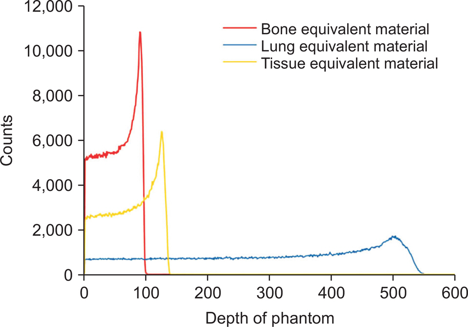
Fig. 3 Prompt gamma depth distribution generated using a 150-MeV proton beam to irradiate bone-, lung-, and tissue-equivalent materials.
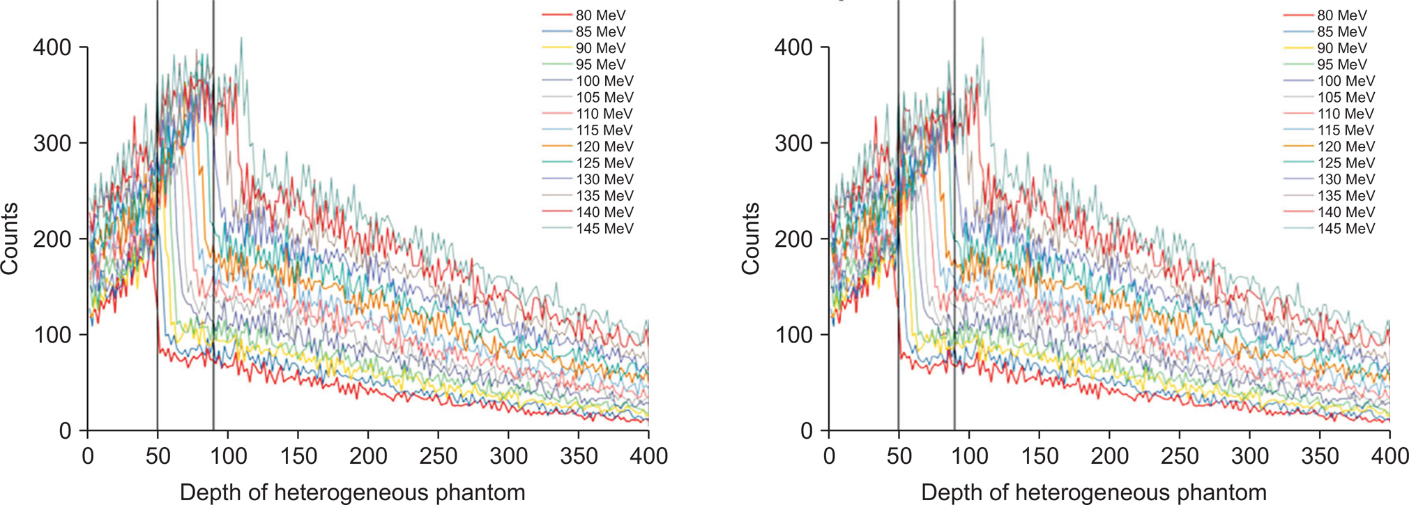
Fig. 4 Variation of prompt-gamma distribution with irradiation proton-beam energy acquired by a multi-slit prompt-gamma camera for the case with bone-equivalent material inside tissue-equivalent material (left side), and prompt-gamma distribution corrected for the influence of the heterogeneous phantom (right side).

Fig. 5 Range results before and after correction of automatic proton-beam range-determination algorithms for the case with bone-equivalent material inside tissue-equivalent material.
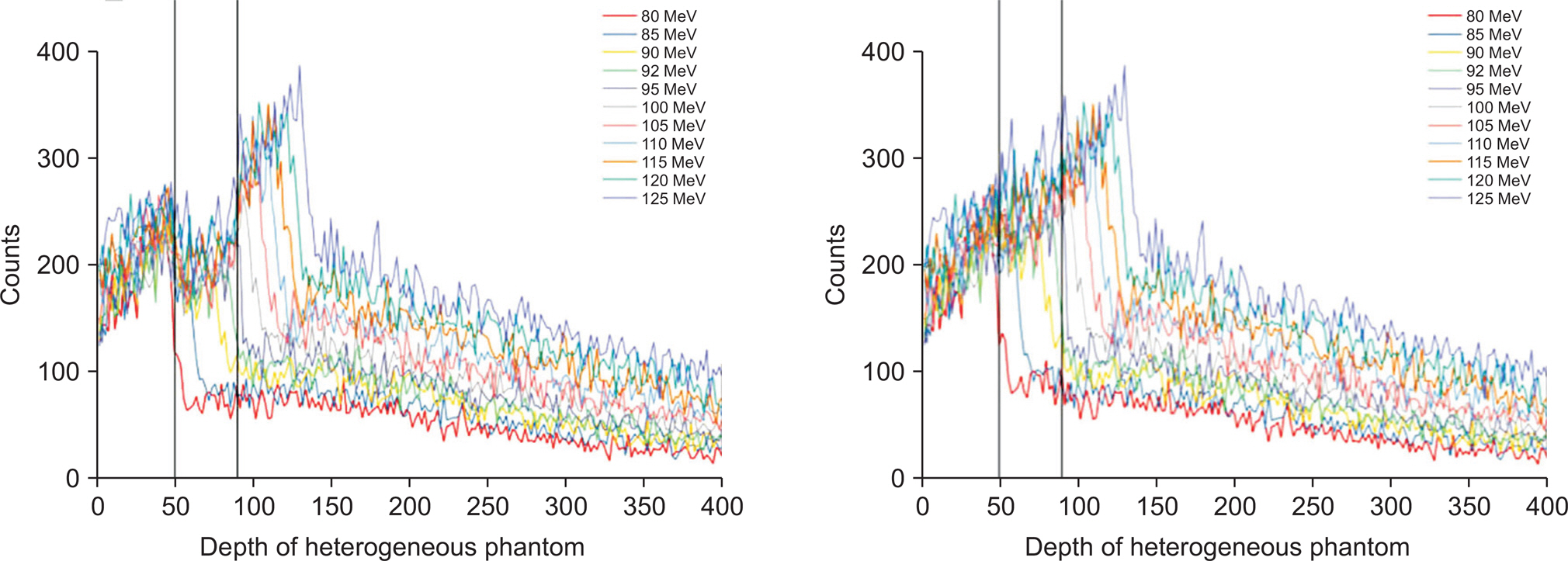
Fig. 6 Variation of prompt-gamma distribution with irradiation proton-beam energy acquired by a multi-slit prompt-gamma camera for the case with lung-equivalent material inside tissue-equivalent material (left side), and prompt-gamma distribution corrected for the influence of the heterogeneous phantom (right side).

Fig. 7 Range results obtained before and after correction of automatic proton-beam range-determination algorithms for the case with lung-equivalent material inside tissue-equivalent material.

Fig. 8 Variation of prompt-gamma distribution with irradiation proton-beam energy acquired by a multi-slit prompt-gamma camera for the case with tissue-equivalent material inside lung-equivalent material (left side), and prompt-gamma distribution corrected for the influence of the heterogeneous phantom (right side).
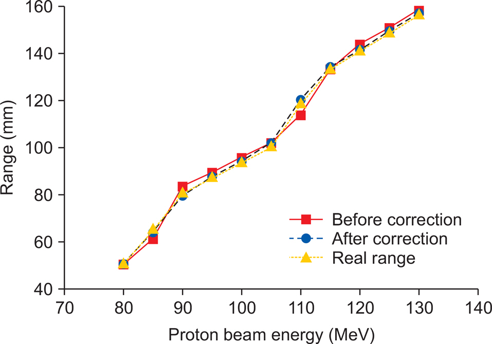
Fig. 9 Range results before and after correction of automatic proton-beam range-determination algorithms for the case with tissue-equivalent material inside lung-equivalent material.
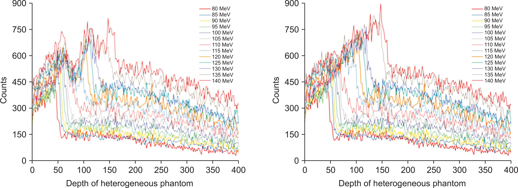
Fig. 10. Variation of prompt-gamma distribution with irradiation proton-beam energy acquired by a multi-slit prompt-gamma camera for the case of a complex heterogeneous phantom (left side), and prompt-gamma distribution corrected for the influence of the heterogeneous phantom (right side).
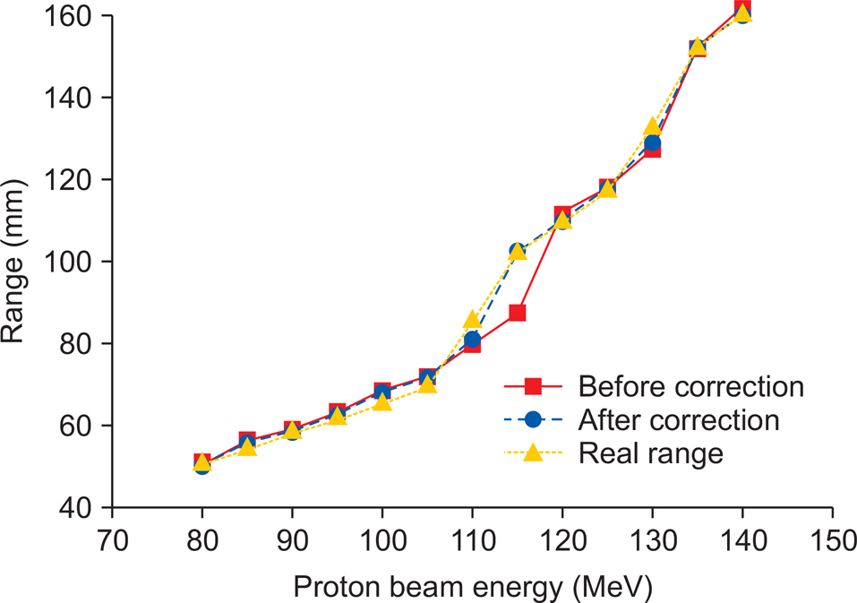
Fig. 11. Range results obtained before and after correction of automatic proton-beam range-determination algorithms for a complex heterogeneous phantom.
Reference
-
1.Paganetti H. Range uncertainties in proton therapy and the role of Monte Carlo simulations. Phys. Med. Biol. 2012. 57:R99–R117.
Article2.Knopf AC., Lomax A. In vivo proton range verification: a review. Phys. Med. Biol. 2012. 58:R131–R160.3.Min CH., Kim CH., Youn MY., Kim JW. Prompt gamma measurements for locating the dose fall-off region in the proton therapy. Appl. Phys. Lett. 2006. 89:183517.
Article4.Park JH., Lee HR., Kim SH, et al. Development of dual-mode signal processing module for multi-slit prompt-gamma camera. Prog. Med. Phys. 2016. 27:37–45.
Article5.Park JH., Lee HR., Kim SH, et al. Development of automated proton beam range verification algorithm for multi-slit prompt-gamma camera. The Korean Association for Radiation Protection Autumn Meeting. Jeju, Korea. 2016.6.ICRP publication 110. Adult reference computational phantoms. 2009. 39(2):7.ICRU Report 46. Photon, Electron, Proton and Neutron International data for Body Tissues. 1992.8.Zhang R., Newhauser W. Calculation of water equivalent thickness of materials of arbitrary density elemental composition and thickness in proton beam irradiation. Phys. Med. Biol. 2009. 54:1383–1395.
Article9.Priegnitz M., Helmbrecht S., Janssens G, et al. Measurement of prompt gamma profiles in heterogeneous targets with a knife-edge slit camera during proton irradiation. Phys. Med. Biol. 2015. 60:4849–4871.10.Schneider W., Bortfeld T., Schlegel W. Correlation between CT numbers and tissue parameters needed for Monte Carlo simulations of clinical dose distributions. Phys. Med. Biol. 2000. 45:459–478.
Article
- Full Text Links
-
- Actions
-
Cited
- CITED
-
- Close
- Share
-
- Similar articles
-
- Development of Two-dimensional Prompt-gamma Measurement System for Verification of Proton Dose Distribution
- Evaluation of Prompt Gamma and Positron Emitter Properties for In-Vivo Dose Verification of Carbon-ion Therapy: A Monte Carlo Study
- Development of Dual-mode Signal Processing Module for Multi-slit Prompt-gamma Camera
- Study on Optimization of Detection System of Prompt Gamma Distribution for Proton Dose Verification
- A Proof-of-Principle Experiment of Hybrid Prompt Gamma-Positron Emission Tomography System for In Vivo Dose Verification in Proton Therapy
