J Korean Assoc Oral Maxillofac Surg.
2017 Dec;43(Suppl 1):S14-S18. 10.5125/jkaoms.2017.43.S1.S14.
Squamous cell carcinoma from oral lichen planus: a case report of a lesion with 28 years of evolution
- Affiliations
-
- 1Department of Dentistry, University of Passo Fundo, Passo Fundo, Brazil.
- 2Department of Dentistry, Federal University of Sergipe, Lagarto, Brazil.
- 3Department of Dentistry, Sagrado Coração University, Bauru, Brazil. pamelalsantos@hotmail.com
- KMID: 2399920
- DOI: http://doi.org/10.5125/jkaoms.2017.43.S1.S14
Abstract
- Lichen planus (LP) is a relatively common mucocutaneous disease with autoimmune etiology. Considering its malignancy potential, it is important to define the correct diagnosis, treatment, and clinical follow-up for patients with LP so that the disease is not diagnosed late, thus hindering the chances of curing the disease. This study aims to describe a clinical case of oral squamous cell carcinoma, potentially originated from LP. The patient is undergoing clinical and histopathological follow-up. A 64-year-old Caucasian male patient presented with a proliferative verrucous lesion on the tongue and sought treatment at the School of Dentistry, University of Passo Fundo (UPF), Passo Fundo, Brazil. He claimed the lesion had been present since 1988, and had been initially diagnoses as "oral lichen planus." The physical exam presented three diagnostic hypotheses: plaque-like oral LP, verrucous carcinoma, and squamous cell carcinoma. After incisional biopsy and histopathological analysis, squamous cell carcinoma was diagnosed, probably originating from oral LP. The case study shows that malignancy from oral LP is possible, which justifies periodic clinical and histopathological follow-up, as well as the elimination of risk factors for carcinoma in patients with oral LP.
MeSH Terms
Figure
-
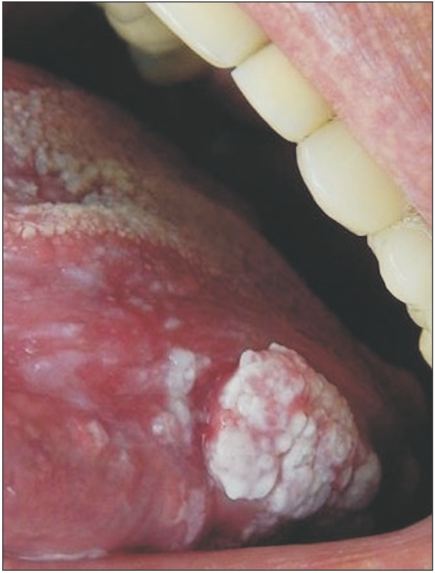
Fig. 1 Initial clinical aspect of the vegetating lesion in the dorsum of the tongue.
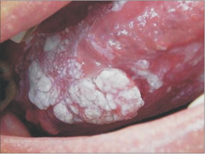
Fig. 2 Initial clinical aspect of the vegetating lesion in the belly of the tongue.
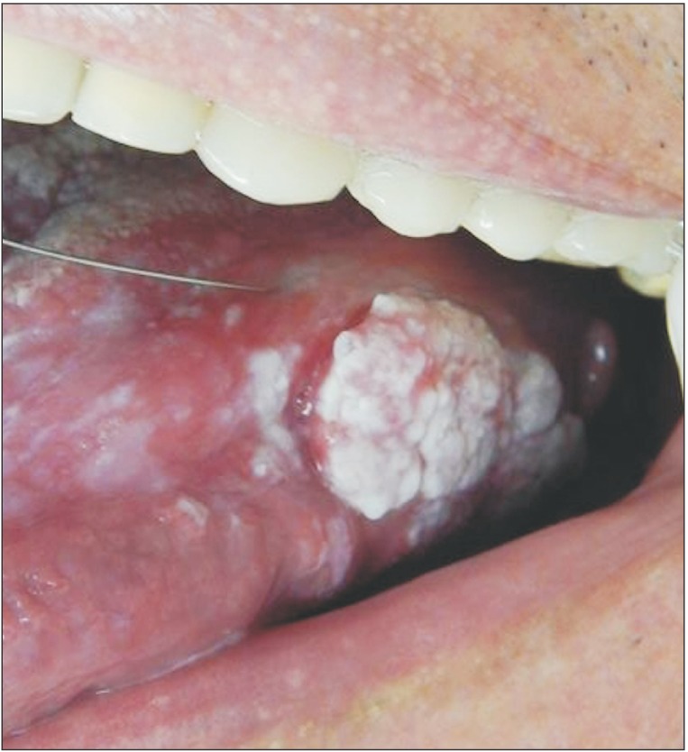
Fig. 3 Anesthetic infiltration before incisional biopsy.
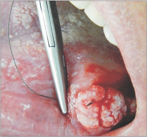
Fig. 4 Collection of a lesion sample with needle and suture thread.
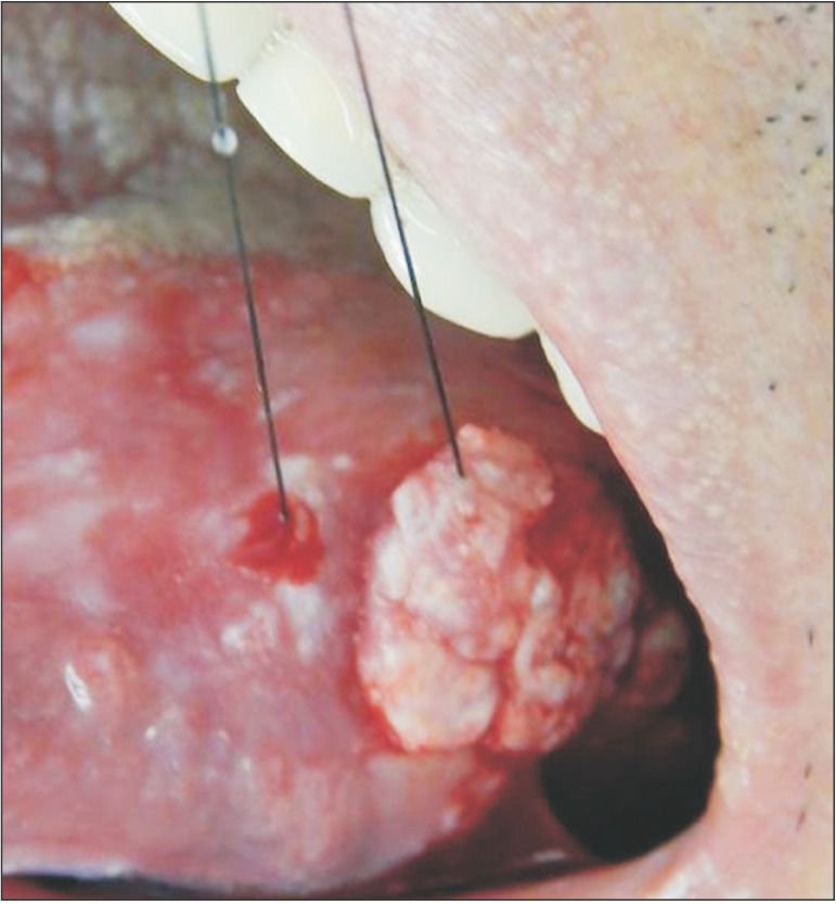
Fig. 5 Lesion with suture thread.
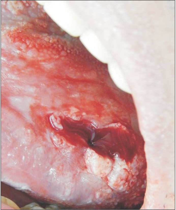
Fig. 6 Scaphoid-shaped surgical site immediately after biopsy.
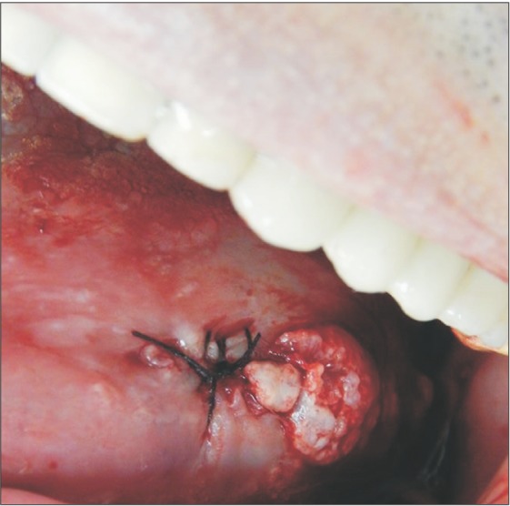
Fig. 7 Suture performed in the incisional biopsy area.
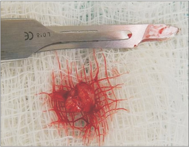
Fig. 8 Aspect of the lesion after incisional biopsy.
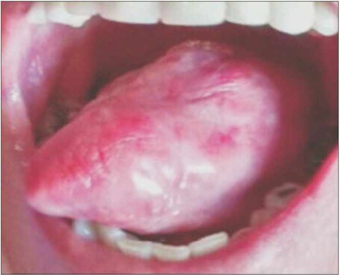
Fig. 9 Aspect of the lesion 2 years after incisional biopsy, 6 chemotherapy sessions, and 35 radiotherapy sessions.
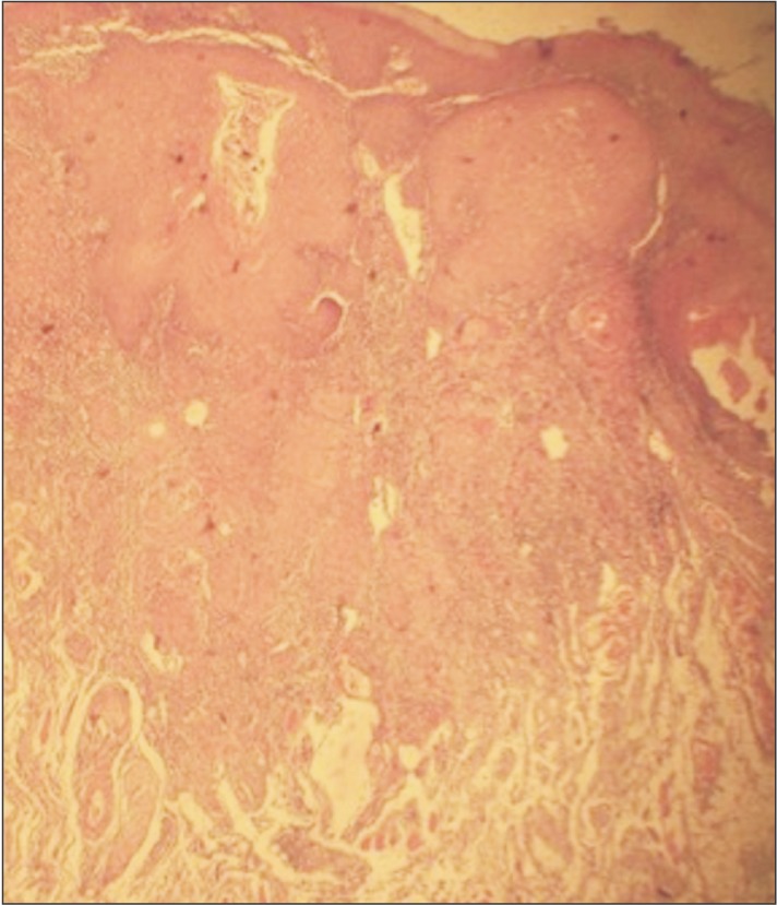
Fig. 10 Grade I squamous cell carcinoma composed of numerous atypical pleomorphic cells and keratin pearls (H&E staining, ×100).
Reference
-
1. Kouhsoltani M, Aghbali A, Shokoohi B, Ahmadzadeh R. Molecular targeting of Her-2/neu protein is not recommended as an adjuvant therapy in oral squamous cell carcinoma and oral lichen planus. Adv Pharm Bull. 2015; 5(Suppl 1):649–652. PMID: 26793611.
Article2. Krupaa RJ, Sankari SL, Masthan KM, Rajesh E. Oral lichen planus: an overview. J Pharm Bioallied Sci. 2015; 7(Suppl 1):S158–S161. PMID: 26015696.
Article3. Santos LCO, Batista OM, Cangussu MCT. Characterization of oral cancer diagnostic delay in the state of Alagoas. Braz J Otorhinolaryngol. 2010; 76:416–422. PMID: 20835525.4. Seoane JM, González-Mosquera A, Velo-Noya J. Oral biopsy in the context of oral cancer and precancer. Av Odontoestomatol. 2008; 24:89–96.5. Bharanidharan R, Dineshkumar T, Raghavendhar K, Kumar AR. Squamous cell carcinoma of the gingiva: a diagnostic enigma. J Oral Maxillofac Pathol. 2015; 19:267.
Article6. de Oliveira JMB, Pinto LO, Lima NGM, de Almeida GCM. Oral cancer: assessment of academic dentistry and nursing knowledge as for the risk factors and diagnostic procedures. Rev Bras Cancerol. 2013; 59:211–218.7. Garcia-Pola MJ, Llorente-Pendás S, González-Garcia M, García-Martín JM. The development of proliferative verrucous leukoplakia in oral lichen planus. A preliminary study. Med Oral Patol Oral Cir Bucal. 2016; 21:e328–e334. PMID: 27031060.
Article8. Budimir V, Richter I, Andabak-Rogulj A, Vučićević-Boras V, Budimir J, Brailo V. Oral lichen planus: retrospective study of 563 Croatian patients. Med Oral Patol Oral Cir Bucal. 2014; 19:e255–e260. PMID: 24608217.9. Shi W, Yang J, Li S, Shan X, Liu X, Hua H, et al. Potential involvement of miR-375 in the premalignant progression of oral squamous cell carcinoma mediated via transcription factor KLF5. Oncotarget. 2015; 6:40172–40185. PMID: 26474386.
Article10. Cabello BT, Sazo BN, Salgado FA, Martínez RB. Squamous cell carcinoma of the lip survival rate. Rev Med Chil. 2015; 143:847–855. PMID: 26361020.11. Mankapure PK, Humbe JG, Mandale MS, Bhavthankar JD. Clinical profile of 108 cases of oral lichen planus. J Oral Sci. 2016; 58:43–47. PMID: 27021539.
Article12. Pedron IG, dos Santos ESR, Aburad A, Tortamano IP, Adde CA. Squamous cell carcinoma: diagnosis and first behavior. J Health Sci Inst. 2006; 24:237–241.13. Selvamani M, Yamunadevi A, Basandi PS, Madhushankari GS. Prevalence of oral squamous cell carcinoma of tongue in and around Davangere, Karnataka, India: a retrospective study over 13 years. J Pharm Bioallied Sci. 2015; 7(Suppl 2):S491–S494. PMID: 26538904.
Article14. Agha-Hosseini F, Mirzaii-Dizgah I. Serum and saliva collagenase-3 (MMP-13) in patients with oral lichen planus and oral squamous cell carcinoma. Med J Islam Repub Iran. 2015; 29:218. PMID: 26478876.15. Oh MS, Kang SH, Kim HJ, Zhenglin Z, Ryu JI, Nam W, et al. Overall five-year survival rate in squamous cell carcinoma of oral cavity. J Korean Assoc Oral Maxillofac Surg. 2009; 35:83–88.16. De Carli JP, Trentin MS, Linden MSS, Bós ÂJG, Pedro REL, Silva SO. Oral squamous cell carcinoma of great extent: protocol diagnosis. Odonto. 2010; 18:67–71.17. Hande AH, Mohite DP, Chaudhary MS, Patel M, Agarwal P, Bohra S. Evidence based demonstration of the concept of ‘field cancerization’ by p53 expression in mirror image biopsies of patients with oral squamous cell carcinoma: an immunohistochemical study. Rom J Morphol Embryol. 2015; 56:1027–1033. PMID: 26662135.18. Brener S, Jeunon FA, Barbosa AA, Grandinetti HAM. Carcinoma de células escamosas bucal: uma revisão de literatura entre o perfil do paciente, estadiamento clínico e tratamento proposto. Rev Bras Cancerol. 2007; 53:63–69.
Article
- Full Text Links
-
- Actions
-
Cited
- CITED
-
- Close
- Share
-
- Similar articles
-
- A Case of Squamous Cell Carcinoma Originating from Oral Lichen Planus
- Invasive Squamous Cell Carcinoma Arising in the Gingival Oral Lichen Planus: Importance of Oral Hygiene
- A Case of Squamous Cell Carcinoma following Erosive Lichen Planus of the Vulva
- Perforating Lichen Nitidus Associated with Oral Lichen Planus
- Localized Lichen Planus on the Lower Lip
