Esophageal Leiomyoma Originating in the Muscularis Propria Layer Resected by Endoscopic Submucosal Dissection
- Affiliations
-
- 1Division of Gastroenterology, Department of Internal Medicine, Ulsan University Hospital, University of Ulsan College of Medicine, Ulsan, Korea. jidmd@uuh.ulsan.kr
- KMID: 2398874
- DOI: http://doi.org/10.4166/kjg.2017.70.5.265
Abstract
- No abstract available.
MeSH Terms
Figure
-
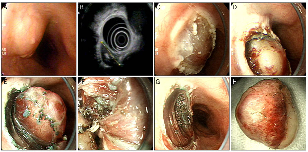
Fig. 1 Endoscopic submucosal dissection process for treating an esophageal subepithelial tumor (SET). (A) A 2.5-cm esophageal SET located 23 cm from the central incisor. (B) An endoscopic ultrasound image showing a homogeneous hypoechoic tumor in the muscularis propria. (C) The covering mucosa of the SET stripped off by a coagulation snare. (D-F) The insulated-tip knife used to separate the tumor from the muscularis propria. (G) The tumor was completely separated from the esophageal wall. (H) Resected specimen, a 2.5×2 cm whitish tumor.
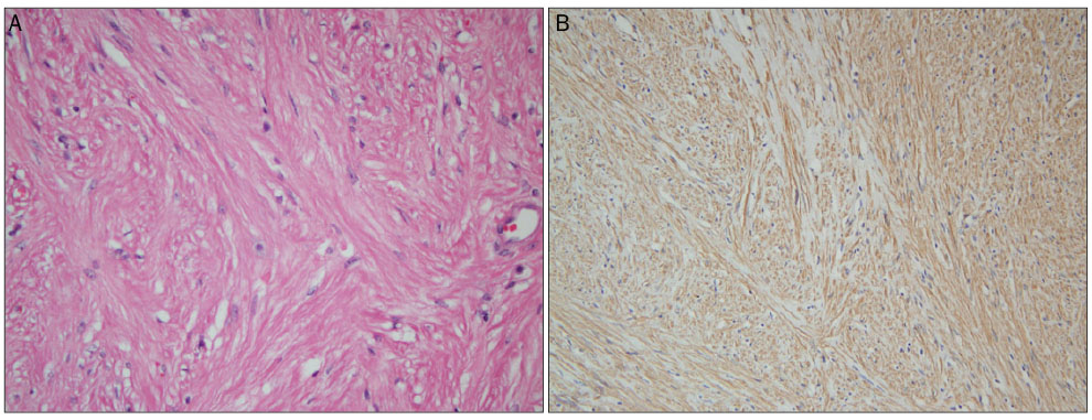
Fig. 2 Microscopic findings of a resected tumor. (A) Microscopically, the tumor was composed of irregularly oriented bundles of smooth muscle cells arranged in a whorl pattern (hemotoxylin and eosin, ×100). (B) Immunohistochemical stain (desmin, ×100) for desmin revealed positivity in the tumor cells.
- Full Text Links
-
- Actions
-
Cited
- CITED
-
- Close
- Share
-
- Similar articles
-
- Endoscopic Submucosal Dissection of a Leiomyoma Originating from the Muscularis Propria of Upper Esophagus
- Submucosal Tunneling Endoscopic Resection of a Leiomyoma Originating from the Muscularis Propria of the Gastric Cardia (with Video)
- A Case of Esophageal Gastrointestinal Stromal Tumor Treated by Endoscopic Submucosal Dissection following an Initial Mucosectomy Using a Transparent Cap
- Endoscopic Characteristics of Upper Gastrointestinal Mesenchymal Tumors Originating from Muscularis Mucosa or Muscularis Propria
- A Submucosal Tumor-Like Recurrence of Early Esophageal Cancer after Endoscopic Submucosal Dissection
