Korean J Gastroenterol.
2013 Oct;62(4):234-237. 10.4166/kjg.2013.62.4.234.
Endoscopic Submucosal Dissection of a Leiomyoma Originating from the Muscularis Propria of Upper Esophagus
- Affiliations
-
- 1Digestive Disease Center and Research Institute, Department of Internal Medicine, Soonchunhyang University College of Medicine, Bucheon, Korea. sjhong@schmc.ac.kr
- 2Department of Pathology, Soonchunhyang University College of Medicine, Bucheon, Korea.
- KMID: 1792742
- DOI: http://doi.org/10.4166/kjg.2013.62.4.234
Abstract
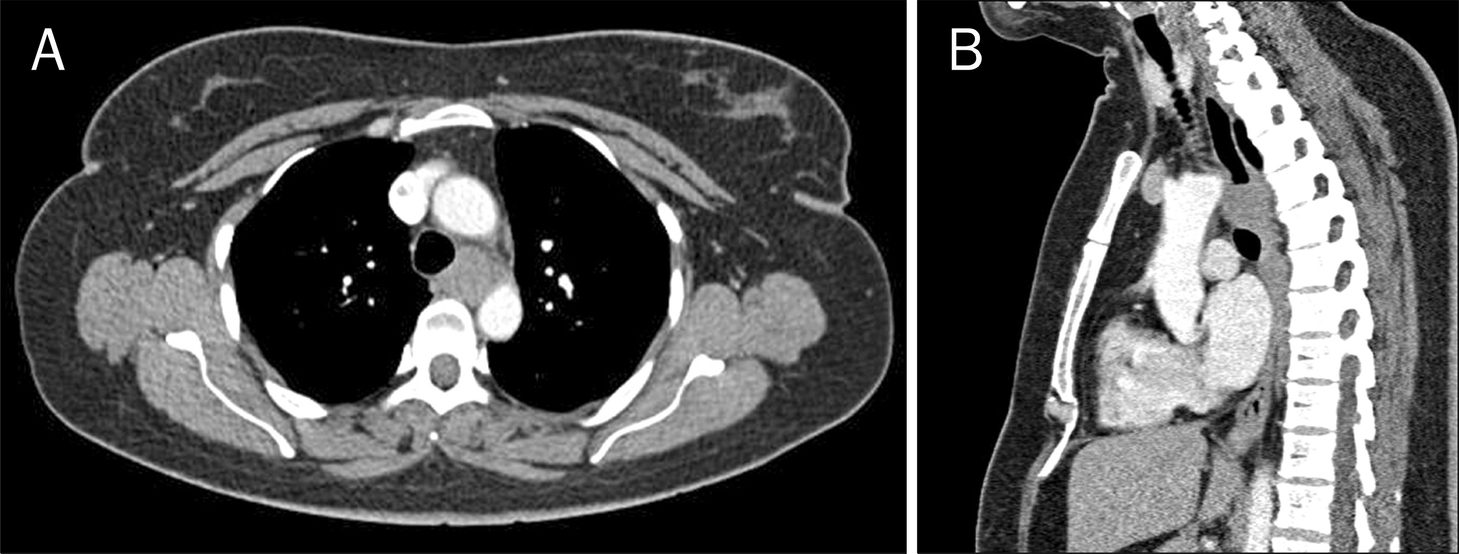
- The technique of endoscopic submucosal dissection is occasionally used for resection of myogenic tumors originating from muscularis mucosa or muscularis propria of stomach and esophagus. However, endoscopic treatments for esophageal myogenic tumors >2 cm have rarely been reported. Herein, we report a case of large leiomyoma originating from muscularis propria in the upper esophagus. A 59-year-old woman presented with dysphagia. Esophagoscopy and endoscopic ultrasonography revealed an esophageal subepithelial tumor which measured 25x20 mm in size, originated from muscularis propria, and was located at 20 cm from the central incisors. The tumor was successfully removed by endoscopic submucosal dissection and there were no complications after en bloc resection. Pathologic examination was compatible with leiomyoma.
MeSH Terms
Figure
Reference
-
References
1. Ono H, Kondo H, Gotoda T, et al. Endoscopic mucosal resection for treatment of early gastric cancer. Gut. 2001; 48:225–229.
Article2. Saito Y, Uraoka T, Matsuda T, et al. Endoscopic treatment of large superficial colorectal tumors: a case series of 200 endoscopic submucosal dissections (with video). Gastrointest Endosc. 2007; 66:966–973.
Article3. Fujishiro M, Yahagi N, Kakushima N, et al. Endoscopic submucosal dissection of esophageal squamous cell neoplasms. Clin Gastroenterol Hepatol. 2006; 4:688–694.
Article4. Honda T, Yamamoto H, Osawa H, et al. Endoscopic submucosal dissection for superficial duodenal neoplasms. Dig Endosc. 2009; 21:270–274.
Article5. Iizuka T, Kikuchi D, Hoteya S, et al. Clinical advantage of endoscopic submucosal dissection over endoscopic mucosal resection for early mesopharyngeal and hypopharyngeal cancers. Endoscopy. 2011; 43:839–843.
Article6. Li QL, Yao LQ, Zhou PH, et al. Submucosal tumors of the esophagogastric junction originating from the muscularis propria layer: a large study of endoscopic submucosal dissection (with video). Gastrointest Endosc. 2012; 75:1153–1158.
Article7. Zhou PH, Yao LQ, Qin XY, et al. Endoscopic full-thickness resection without laparoscopic assistance for gastric submucosal tumors originated from the muscularis propria. Surg Endosc. 2011; 25:2026–2931.
Article8. Lee IL, Lin PY, Tung SY, Shen CH, Wei KL, Wu CS. Endoscopic submucosal dissection for the treatment of intraluminal gastric subepithelial tumors originating from the muscularis propria layer. Endoscopy. 2006; 38:1024–1028.
Article9. Liu BR, Song JT, Qu B, Wen JF, Yin JB, Liu W. Endoscopic muscularis dissection for upper gastrointestinal subepithelial tumors originating from the muscularis propria. Surg Endosc. 2012; 26:3141–3148.
Article10. Shi Q, Zhong YS, Yao LQ, Zhou PH, Xu MD, Wang P. Endoscopic submucosal dissection for treatment of esophageal submucosal tumors originating from the muscularis propria layer. Gastrointest Endosc. 2011; 74:1194–1200.
Article11. Gong W, Xiong Y, Zhi F, Liu S, Wang A, Jiang B. Preliminary experience of endoscopic submucosal tunnel dissection for upper gastrointestinal submucosal tumors. Endoscopy. 2012; 44:231–235.
Article12. Seremetis MG, Lyons WS, deGuzman VC, Peabody JW Jr. Leiomyomata of the esophagus. An analysis of 838 cases. Cancer. 1976; 38:2166–2177.13. Kabuto T, Taniguchi K, Iwanaga T, Terasawa T, Tateishi R, Taniguchi H. Diffuse leiomyomatosis of the esophagus. Dig Dis Sci. 1980; 25:388–391.
Article14. Gallinger S, Steinhardt MI, Goldberg M. Giant leiomyoma of the esophagus. Am J Gastroenterol. 1983; 78:708–711.15. Preda F, Alloisio M, Lequaglie C, Ongari M, Ravasi G. Leiomyoma of the esophagus. Tumori. 1986; 72:503–506.
Article16. Lee LS, Singhal S, Brinster CJ, et al. Current management of esophageal leiomyoma. J Am Coll Surg. 2004; 198:136–146.
Article17. Roviaro GC, Maciocco M, Varoli F, Rebuffat C, Vergani C, Scarduelli A. Videothoracoscopic treatment of oesophageal leiomyoma. Thorax. 1998; 53:190–192.
Article18. Hu B, Mou Y, Yi H, et al. Endoscopic enucleation of large esophageal leiomyomas. Gastrointest Endosc. 2011; 74:928–931.
Article19. Yoon HJ, Ryu CB, Na HS, et al. The usefulness of endoscopic sub-tumoral dissection for en-bloc resection of upper gastrointestinal submucosal tumor. Korean J Gastrointest Endosc. 2008; 36:193–199.
- Full Text Links
- Actions
-
Cited
- CITED
-
- Close
- Share
- Similar articles
-
- Submucosal Tunneling Endoscopic Resection of a Leiomyoma Originating from the Muscularis Propria of the Gastric Cardia (with Video)
- Endoscopic Characteristics of Upper Gastrointestinal Mesenchymal Tumors Originating from Muscularis Mucosa or Muscularis Propria
- Esophageal Leiomyoma Originating in the Muscularis Propria Layer Resected by Endoscopic Submucosal Dissection
- A Case of Esophageal Gastrointestinal Stromal Tumor Treated by Endoscopic Submucosal Dissection following an Initial Mucosectomy Using a Transparent Cap
- Endoscopic Submucosal Tunnel Dissection for Upper Gastrointestinal Submucosal Tumors Originating from the Muscularis Propria Layer: A Single-Center Study


