J Vet Sci.
2017 Dec;18(4):563-565. 10.4142/jvs.2017.18.4.563.
Mineralized deposits in the uterus of a pig without pregnancy loss
- Affiliations
-
- 1Department of Theriogenology and Biotechnology, Research Institute for Veterinary Science, College of Veterinary Medicine, Seoul National University, Seoul 08826, Korea. bclee@snu.ac.kr
- 2Laboratory of Histology and Molecular Pathogenesis, College of Veterinary Medicine, Kangwon National University, Chuncheon 24341, Korea.
- 3Laboratory of Theriogenology, College of Veterinary Medicine, Chungnam National University, Daejeon 34134, Korea.
- KMID: 2398519
- DOI: http://doi.org/10.4142/jvs.2017.18.4.563
Abstract
- Herein, we describe a case of uterine calcification in the uterus of a pig without pregnancy loss. The recipient underwent cloned embryo transfer and Cesarean section for safe delivery of cloned piglets. During the Cesarean section, 4 white, star-like, (2 × 2 × 2) cm, calcified structures were found within the endometrial cavity. Despite dystrophic calcification around the placenta, healthy cloned piglets were produced successfully. To our knowledge, this is the first reported case of dystrophic calcification occurring within the uterus in a pregnant pig.
Figure
-
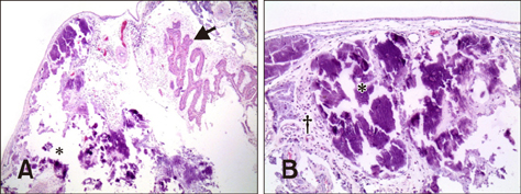
Fig. 1 Histopathological images of abnormal tissue attached to the maternal uterus. (A) The placenta (arrow) and a broad area of dystrophic calcification (*). (B) Calcified structures are observed in uterine tissue. Note the broad area of mineralized deposits (*) and infiltration of inflammatory cells (†), mainly histiocytes. 40× (A), 100× (B).
Reference
-
1. Alpert LC, Haufrect EJ, Schwartz MR. Uterine lithiasis. Am J Surg Pathol. 1990; 14:1071–1075.
Article2. Cayuela E, Perez-Medina T, Vilanova J, Alejo M, Cañadas P. True osseous metaplasia of the endometrium: the bone is not from a fetus. Fertil Steril. 2009; 91:1293.e1–1293.e4.
Article3. Cicinelli E, Stanziano A, Parisi C, Marinaccio M, Causio F. Hysteroscopic diagnosis and treatment of endocervical ossification: a case report. J Minim Invasive Gynecol. 2005; 12:159–161.
Article4. Feyles V, Moyana TN, Pierson RA. Recurrent pregnancy loss associated with endometrial hyperechoic areas (endometrial calcifications): a case report and review of the literature. Clin Exp Obstet Gynecol. 2000; 27:5–8.5. Iwasaki M, Oliveira CA. Uterine lithiasis in a dog. Aust Vet J. 1991; 68:73–74.
Article6. Jin JX, Lee S, Khoirinaya C, Oh A, Kim GA, Lee BC. Supplementation with spermine during in vitro maturation of porcine oocytes improves early embryonic development after parthenogenetic activation and somatic cell nuclear transfer. J Anim Sci. 2016; 94:963–970.
Article7. Misra RP. Calcium and disease: molecular determinants of calcium crystal deposition diseases. Cell Mol Life Sci. 2000; 57:421–428.
Article8. Onderoglu LS, Yarali H, Gultekin M, Katlan D. Endometrial osseous metaplasia: an evolving cause of secondary infertility. Fertil Steril. 2008; 90:2013.e9–2013.e11.
Article9. Pereira MC, Vaz MM, Miranda SP, Araújo SR, Menezes DB, das Chagas Medeiros F. Uterine cavity calcifications: a report of 7 cases and a systematic literature review. J Minim Invasive Gynecol. 2014; 21:346–352.
Article10. Ryan LM, Cheung HS. The role of crystals in osteoarthritis. Rheum Dis Clin North Am. 1999; 25:257–267.
Article11. Silva EG, Deavers MT, Parlow AF, Gershenson DM, Malpica A. Calcifications in ovary and endometrium and their neoplasms. Mod Pathol. 2003; 16:219–222.
Article12. Varma VA, Kim KM. Placental calcification: ultrastructural and X-ray microanalytic studies. Scan Electron Microsc. 1985; 1567–1572.
- Full Text Links
-
- Actions
-
Cited
- CITED
-
- Close
- Share
-
- Similar articles
-
- A Case of Pregnancy in a Non-communicating Rudimentary Horn of Unicornuate Uterus After Intrauterine Insemination
- A Case of Ruptured Cornual Pregnancy in Adenomyosis Uterus at 29 Weeks' Gestation
- Tubal Pregnancy in a Unicornuate Uterus with Rudimentary Horn
- Unruptured Cornual Pregnancy: A Case Report
- A successful term pregnancy in a patient with unicornuate uterus
