3-dimensional reconstruction of mandibular canal at the interforaminal region using micro-computed tomography in Korean
- Affiliations
-
- 1Department of Anatomy, College of Medicine, Chosun University, Gwangju, Republic of Korea.
- 2Department of Oral Anatomy, College of Dentistry, Chosun University, Gwangju, Republic of Korea. sky@chosun.ac.kr
- 3Department of Dental Prosthetics, College of Dentistry, Chosun University, Gwangju, Republic of Korea.
- KMID: 2398052
- DOI: http://doi.org/10.4047/jap.2017.9.6.470
Abstract
- PURPOSE
The purpose of this study was to identify the complex course of the mandibular canal using 3D reconstruction of microCT images and to provide the diagram for clinicians to help them understand at the interforaminal region in Korean.
MATERIALS AND METHODS
Twenty-six hemimandibles obtained from cadavers were examined using microCT, and the images were reconstructed. At both the midpoint of mental foramen and the tip of anterior loop, the bucco-lingual position, the height from the mandibular inferior border, the horizontal distance between two points, and position relative to tooth site on the mandibular canal were measured. The angle that the mental canal diverges from the mandibular canal was measured in posteriorsuperior and lateral-superior direction.
RESULTS
The buccal distance from the mandibular canal was significantly much shorter than lingual distance at both the mental foramen and the tip of anterior loop. The mandibular canal at the tip of anterior loop was significantly located closer to buccal side and higher than at the mental foramen. And the mental canal most commonly diverged from the mandibular canal below the first premolar by approximately 50° posterior-superior and 41° lateral-superior direction, which had with a mean length of 5.19 mm in front of the mental foramen, and exited to the mental foramen below the second premolar.
CONCLUSION
These results suggest that it could form a hazardous tetrahedron space at the interforaminal region, thus, the clinician need to pay attention to the width of a premolar tooth from the mental foramen during dental implant placement.
Figure
-
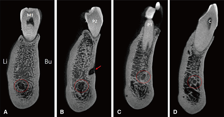
Fig. 1 Serial images (coronal views) of the mandibular canal obtained using microCT scanning. The dashed circle indicates the course of the mandibular canal at each tooth site. The arrow indicates the opening (mental foramen) of mental canal. (A) M1, first molar; (B) P2, second premolar; (C) P1, first premolar; (D) C, canine. Bu, buccal side; Li, lingual side.

Fig. 2 Three-dimensional reconstructed images of the mandibular canal. (A – C, capital letter of top line) Lateral views showing the 3 types of the mandibular canal according to the divergent in the posterior-superior direction: type 1 (A), type 2 (B), and type 3 (C). (a – c, small letter of bottom line) Anteroposterior views of the mandibular canal corresponding to the images in A – C, respectively.
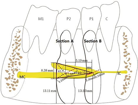
Fig. 3 Schematic drawing shows the major course of the mandibular canal which forms a hazardous tetrahedron space at the interforaminal region. The measurement was performed in the reference position where is at both the midpoint of mental foramen (Section A) and the tip of anterior loop (Section B). The data represent mean values of each measurement items. MC, mandibular canal; mc, mental canal; IC, incisive canal.
Reference
-
1. Liang XH, Kim YM, Cho IH. Residual bone height measured by panoramic radiography in older edentulous Korean patients. J Adv Prosthodont. 2014; 6:53–59.2. Feine JS, Carlsson GE, Awad MA, Chehade A, Duncan WJ, Gizani S, Head T, Lund JP, MacEntee M, Mericske-Stern R, Mojon P, Morais J, Naert I, Payne AG, Penrod J, Stoker GT Jr, Tawse-Smith A, Taylor TD, Thomason JM, Thomson WM, Wismeijer D. The McGill Consensus Statement on Overdentures. Montreal, Quebec, Canada. May 24-25, 2002. Int J Prosthodont. 2002; 15:413–414.3. Watanabe H, Mohammad Abdul M, Kurabayashi T, Aoki H. Mandible size and morphology determined with CT on a premise of dental implant operation. Surg Radiol Anat. 2010; 32:343–349.4. Kim MK. Head and neck anatomy. 5th ed. Seoul: Dental & Medical Publishing;2011. p. 86.5. Li X, Jin ZK, Zhao H, Yang K, Duan JM, Wang WJ. The prevalence, length and position of the anterior loop of the inferior alveolar nerve in Chinese, assessed by spiral computed tomography. Surg Radiol Anat. 2013; 35:823–830.6. Juodzbalys G, Wang HL, Sabalys G. Anatomy of mandibular vital structures. part II: Mandibular incisive canal, mental foramen and associated neurovascular bundles in relation with dental implantology. J Oral Maxillofac Res. 2010; 1:e3.7. Hwang K, Lee WJ, Song YB, Chung IH. Vulnerability of the inferior alveolar nerve and mental nerve during genioplasty: an anatomic study. J Craniofac Surg. 2005; 16:10–14.8. Kim ST, Hu KS, Song WC, Kang MK, Park HD, Kim HJ. Location of the mandibular canal and the topography of its neurovascular structures. J Craniofac Surg. 2009; 20:936–939.9. Apostolakis D, Brown JE. The anterior loop of the inferior alveolar nerve: prevalence, measurement of its length and a recommendation for interforaminal implant installation based on cone beam CT imaging. Clin Oral Implants Res. 2012; 23:1022–1030.10. Kim HJ, Yu SK, Lee MH, Lee HJ, Kim HJ, Chung CH. Cortical and cancellous bone thickness on the anterior region of alveolar bone in Korean: a study of dentate human cadavers. J Adv Prosthodont. 2012; 4:146–152.11. Yu SK, Lee MH, Jeon YH, Chung YY, Kim HJ. Anatomical configuration of the inferior alveolar neurovascular bundle: a histomorphometric analysis. Surg Radiol Anat. 2016; 38:195–201.12. Jacobs R, Mraiwa N, Van Steenberghe D, Sanderink G, Quirynen M. Appearance of the mandibular incisive canal on panoramic radiographs. Surg Radiol Anat. 2004; 26:329–333.13. Mardinger O, Chaushu G, Arensburg B, Taicher S, Kaffe I. Anterior loop of the mental canal: an anatomical-radiologic study. Implant Dent. 2000; 9:120–125.14. Lee MH, Kim HJ, Kim DK, Yu SK. Histologic features and fascicular arrangement of the inferior alveolar nerve. Arch Oral Biol. 2015; 60:1736–1741.15. Uchida Y, Noguchi N, Goto M, Yamashita Y, Hanihara T, Takamori H, Sato I, Kawai T, Yosue T. Measurement of anterior loop length for the mandibular canal and diameter of the mandibular incisive canal to avoid nerve damage when installing endosseous implants in the interforaminal region: a second attempt introducing cone beam computed tomography. J Oral Maxillofac Surg. 2009; 67:744–750.16. Choi DY, Sun KH, Won SY, Lee JG, Hu KS, Kim KD, Kim HJ. Trabecular bone ratio of the mandibular condyle according to the presence of teeth: a micro-CT study. Surg Radiol Anat. 2012; 34:519–526.17. Engelke K, Song SM, Glüer CC, Genant HK. A digital model of trabecular bone. J Bone Miner Res. 1996; 11:480–489.18. Song WC, Jo DI, Lee JY, Kim JN, Hur MS, Hu KS, Kim HJ, Shin C, Koh KS. Microanatomy of the incisive canal using three-dimensional reconstruction of microCT images: an ex vivo study. Oral Surg Oral Med Oral Pathol Oral Radiol Endod. 2009; 108:583–590.19. Yu SK, Kim S, Kang SG, Kim JH, Lim KO, Hwang SI, Kim HJ. Morphological assessment of the anterior loop of the mandibular canal in Koreans. Anat Cell Biol. 2015; 48:75–80.20. Moiseiwitsch JR. Position of the mental foramen in a North American, white population. Oral Surg Oral Med Oral Pathol Oral Radiol Endod. 1998; 85:457–460.21. Greenstein G, Tarnow D. The mental foramen and nerve: clinical and anatomical factors related to dental implant placement: a literature review. J Periodontol. 2006; 77:1933–1943.22. Choi Y, Jung UW, Um YJ, Kim ST, Hu KS, Kim CS, Kim KD, Chai JK, Choi SH. Running pattern of the mandibular canal in Korean population, by 3-dimensional reconstructed computed tomographic imaging analysis. Implantology. 2009; 13:152–161.23. Kim MS, Kim SH, Kim HJ, Kim HJ, Park BG, Park BS, Park JT, Park JC, Bae YC, Yu SK, Lee YH, Jung HS, Cho SW, Cho US, Heo GS. Dental anatomy and morphology. 3rd ed. Seoul: DaehanNarae Publishing Inc.;2016. p. 163–176.24. de Freitas V, Madeira MC, Pinto CT, Zorzetto NL. Direction of the mental canal in human mandibles. Aust Dent J. 1976; 21:338–340.25. Krekmanov L, Kahn M, Rangert B, Lindström H. Tilting of posterior mandibular and maxillary implants for improved prosthesis support. Int J Oral Maxillofac Implants. 2000; 15:405–414.26. Hu KS, Yun HS, Hur MS, Kwon HJ, Abe S, Kim HJ. Branching patterns and intraosseous course of the mental nerve. J Oral Maxillofac Surg. 2007; 65:2288–2294.27. Mraiwa N, Jacobs R, Moerman P, Lambrichts I, van Steenberghe D, Quirynen M. Presence and course of the incisive canal in the human mandibular interforaminal region: two-dimensional imaging versus anatomical observations. Surg Radiol Anat. 2003; 25:416–423.
- Full Text Links
-
- Actions
-
Cited
- CITED
-
- Close
- Share
-
- Similar articles
-
- Radiographic evaluation of the course and visibility of the mandibular canal
- Analysis and evaluation of relative positions of mandibular third molar and mandibular canal impacts
- Morphological assessment of the anterior loop of the mandibular canal in Koreans
- Evaluation of canal preparation with Ni-Ti rotary files by micro computed tomography
- Evaluation of mesial root canal configuration of mandibular first molars using micro-computed tomography
